
home > treatments > brain biopsy
Brain biopsy
Taking a tissue sample from the brain
What is a brain biopsy?
In a brain biopsy, a small piece of tissue is taken from an abnormality in the brain so that it can be examined under the microscope and with molecular tests. The aim is not to remove the abnormality, but to find out exactly what it is. That knowledge determines further treatment.
Why is a biopsy proposed?
An MRI scan shows that there is an abnormality, but often not with certainty what it is. A brain tumour, inflammation, infection or metastasis can look very similar on imaging, while the treatment is completely different. In series of deep lesions, the diagnosis based on MRI alone turns out not to match the tissue examination in about 15 to 20 in 100 cases.
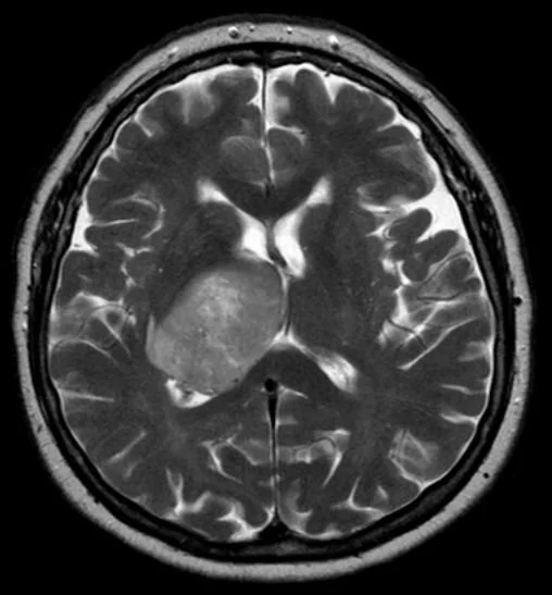
A biopsy is mainly proposed when:
- a tissue diagnosis is needed and complete removal is not (yet) advisable;
- the abnormality cannot be safely removed completely, for example because it lies deep or in an area that controls important functions (speech, movement, vision);
- there are several abnormalities and their origin is unclear;
- the suspected diagnosis is a condition that is not treated surgically, such as lymphoma or an inflammation or infection, and treatment can only start after confirmation;
- the patient's condition is such that a major operation would be too demanding.
If the abnormality can be removed safely, complete removal is usually preferable to a biopsy: it provides both the diagnosis and the treatment at the same time. If we suspect lymphoma, we ask that steroids (dexamethasone) be postponed until after the biopsy if possible: steroids make lymphoma cells disappear quickly and can make the sample unusable.
How is the procedure carried out?
The most commonly used technique is stereotactic needle biopsy with neuronavigation: a thin needle, guided by the computer, is passed through a small hole in the skull into the abnormality.
Neuronavigation. Neuronavigation works like a GPS for the brain. Before the procedure, an MRI is done; in the operating theatre, the computer links those images to your head. A camera tracks the instruments using small reflective spheres, so that the surgeon can see on the screen at any moment where the needle tip is in your brain, with an accuracy of a few millimetres. Dr Lavrysen works with a latest-generation navigation system, which allows the trajectory to be adjusted during the procedure if necessary.

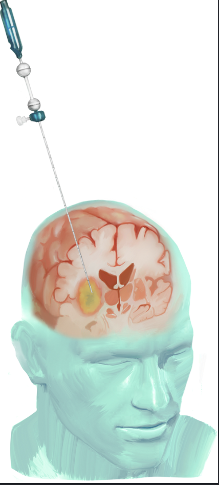
- Planning: based on a recent MRI, a safe trajectory to the abnormality is calculated with computer navigation, steering clear of blood vessels and important brain areas.
- Procedure: under general anaesthesia, or sometimes local anaesthesia with light sedation, a hole of a few millimetres is made in the skull through a small skin incision. A fine needle is passed along the planned trajectory into the abnormality, where a few small tissue samples are taken, often at different depths.
- Check: the tissue is already briefly examined by the pathologist during the procedure to check whether the sample is representative. If not, additional samples are taken. This roughly halves the number of biopsies without a diagnosis. The skin is closed with a few stitches or staples.
The procedure usually takes less than an hour. It is usually followed by a check-up scan and a hospital stay of one to two nights.
For an abnormality just below the surface of the brain, an open biopsy can also be done through a slightly larger opening, with the tissue removed under direct vision using the microscope.
What are the possible risks?
A brain biopsy is a minor procedure, but it is not without risk. The figures below come from large series and review studies of stereotactic biopsies.
- Bleeding at the biopsy site: a small bleed that is only visible on the check-up scan and causes no symptoms occurs in about 15 to 20 in 100. A bleed that causes symptoms (weakness, speech disturbance, drowsiness) occurs in about 1 in 20; this loss of function is usually temporary.
- Permanent loss of function due to a bleed or damage to brain tissue: about 1 to 2 in 100.
- Death as a result of the procedure, almost always due to a major bleed: about 1 in 100 in older series, less in recent ones.
- Epileptic seizure after the procedure, in a small number of patients.
- Wound infection: rare.
- No conclusive diagnosis: in about 1 in 15, the sample is too small or not representative, and a second biopsy or a different approach is needed.
The risk depends on the location of the abnormality (deep lesions bleed more often and more often fail to yield a diagnosis), on the suspected type (lymphomas and tumours with a rich blood supply bleed more often), on blood thinners and on your general condition. We discuss this beforehand.
Recovery and result
Most patients are home again after one to two days and quickly resume their normal activities. Headache and fatigue may last a few days. The stitches are removed after about ten days.
A preliminary result is often available after a few days; the final result of the tissue examination follows after one to two weeks. For tumours, molecular testing is usually also done (for example for IDH mutation, 1p/19q or MGMT), which can take longer but is necessary under the current classification to determine the type of tumour and the best treatment. The result is discussed at a multidisciplinary oncology meeting and then with you.
Frequently asked questions
Why isn't the abnormality removed completely straight away?
Because that cannot always be done safely, or because the suspected diagnosis is a condition that is not treated with surgery. A biopsy then gives the certainty needed to start the right treatment, with a smaller risk.
Will I feel anything during the procedure?
No, the procedure is done under anaesthesia. Brain tissue itself has no sense of pain. Afterwards the wound may be a little tender and headache is possible.
Will my head be shaved?
Only a small area around the incision, usually a few centimetres.
Can the needle hit a blood vessel?
The trajectory is planned in advance so that blood vessels are avoided. Bleeding remains the main risk, but is usually small and without consequences; a bleed with symptoms occurs in about 1 in 20.
What if the sample does not provide a diagnosis?
That happens in about 1 in 15. We then discuss whether a second biopsy, a different technique or further monitoring with imaging makes most sense.
Can a biopsy make the tumour spread?
With brain tumours, contrary to what is sometimes feared, this is not a relevant risk.
How long until I know the result?
A preliminary result is often available after a few days, the final one after one to two weeks. Molecular tests can take a few extra weeks.