
home > treatments > meningiomas
Meningiomas
Benign growth of the brain's membranes
What is a meningioma?
A meningioma is a growth that arises from the meninges, the protective layers between the skull and the brain. So it does not grow from the brain tissue itself, but pushes the brain aside from the outside.
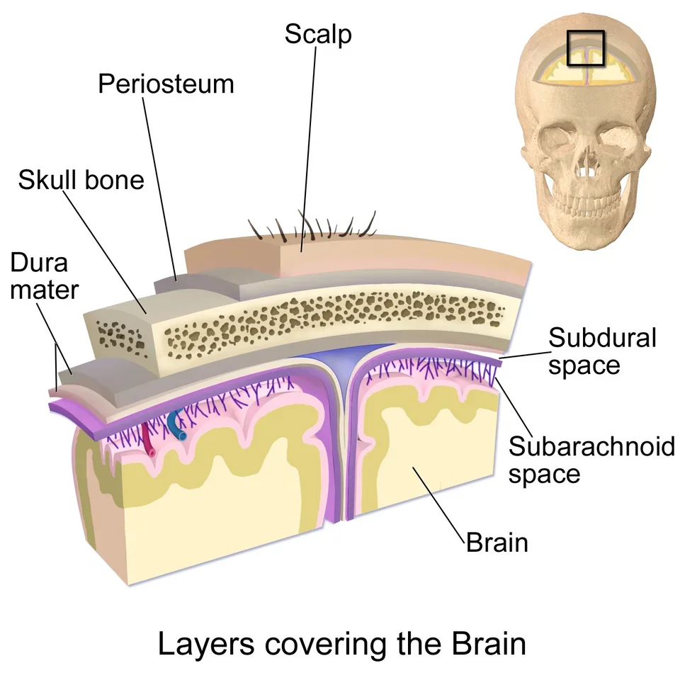
It is the most common brain tumour in adults. Meningiomas occur more often in women and at an older age. About nine in ten are benign (grade 1): they grow slowly and do not spread. A smaller proportion grows faster and comes back more often (grade 2), and a rare minority behaves malignantly (grade 3).
What symptoms does a meningioma cause?
Because meningiomas grow slowly, they often cause no symptoms for a long time. Many meningiomas are discovered by chance on a scan done for another reason.
Possible symptoms depend on the location and size:
- Headache, especially if it is new or gradually getting worse
- Epileptic seizures
- Weakness or numbness in an arm or leg
- Problems with vision, smell or hearing
- Changes in memory, behaviour or concentration
Warning signs: a first epileptic seizure, sudden loss of strength or speech disturbance, or rapidly worsening headache with vomiting or drowsiness. In that case, contact a doctor or the emergency department immediately.
How is the diagnosis made?
An MRI scan with contrast is the most important investigation. Meningiomas have a very recognisable appearance on it, so the diagnosis can usually be made with high probability without examining tissue. Sometimes a CT scan is added to assess the skull or calcifications in the tumour.
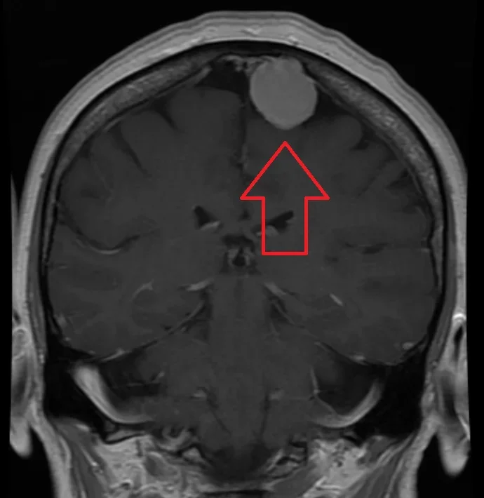
Only microscopic examination of the tissue after an operation gives complete certainty about the type and grade.
Does a meningioma always need treatment?
No. This is an important point: many meningiomas never need to be treated.
A small meningioma without symptoms, especially in older patients, is often simply monitored with an MRI after 6 to 12 months and then at longer intervals. If it remains stable, continued follow-up is sufficient.
Treatment is considered in case of:
- Symptoms caused by the meningioma
- Clear growth during follow-up
- Swelling of the surrounding brain tissue on the scan
- A size or location where future problems can be expected, especially in younger patients
- Doubt about the diagnosis
What treatments are available?
An operation is the main treatment when intervention is needed. The aim is to remove the tumour as completely as possible, together with its attachment to the membrane, without damaging the brain or important nerves and blood vessels.
The procedure is performed under general anaesthesia using the operating microscope, with computer navigation to locate the tumour precisely. For tumours close to important functions, nerve or brain function is monitored electrically during the operation.
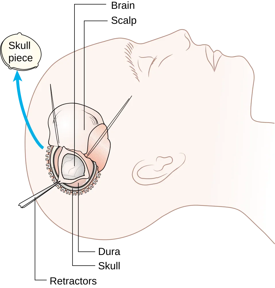
Sometimes a small part is deliberately left behind because complete removal would be too risky, for example when the tumour is adherent to an important blood vessel or cranial nerve. That remnant is then monitored or irradiated. The hospital stay usually lasts from a few days to a week.
Radiation (radiotherapy or radiosurgery) is an alternative or addition: for a remnant after surgery, for regrowth, for tumours in hard-to-reach places, or for patients for whom an operation would be too demanding. Radiation does not remove the tumour, but slows its growth.
There is currently no medication with proven effectiveness against meningiomas.
What are the possible risks of an operation?
The risks depend strongly on the location and size of the tumour. A small meningioma at the top of the skull can usually be removed safely; a tumour at the skull base, between nerves and blood vessels, is more complex. Possible complications:
- Bleeding or swelling of the brain tissue, with temporary or, rarely, permanent loss of function
- Epileptic seizures
- Loss of function of a cranial nerve with tumours at the skull base (face, hearing, eye movements, swallowing)
- Wound infection or leakage of cerebrospinal fluid
- General risks of an operation: thrombosis, pneumonia
We discuss beforehand which risks apply in your situation and how they compare to the risk of waiting.
Recovery and follow-up
Most patients feel well again after a few weeks; full recovery can take a few months. Fatigue and headache are normal during that period. Driving is not allowed for some time after brain surgery; we will discuss with you when you may drive again.
Even after complete removal, follow-up with MRI remains necessary, usually annually and then at longer intervals, because a meningioma can come back after years. For a grade 2 or 3 meningioma, follow-up is more intensive and additional radiotherapy afterwards is often advised.
Frequently asked questions
Is a meningioma cancer?
Usually not. The vast majority are benign, grow slowly and do not spread. A minority behave more aggressively; tissue examination after an operation determines the grade.
My meningioma was found by chance and I have no symptoms. Does it need to be removed?
Often not. A follow-up MRI after 6 to 12 months shows whether it is growing. If it remains stable, follow-up is sufficient.
Can it come back after an operation?
Yes, it can, even after years. The chance is small after complete removal of a benign meningioma and greater when there is a remnant or a higher grade. That is why follow-up with MRI remains important.
Can a meningioma be treated with medication?
No, there is currently no medicine that makes a meningioma shrink. Hormonal factors may play a role in its growth; so discuss any hormone use with your doctor.
Will I be different after the operation?
For most patients, no. Temporary fatigue, headache or concentration problems are normal in the first weeks. Permanent changes are rare and depend on the location of the tumour.
Can I drive after the operation?
Not immediately. The waiting period depends on the procedure and on whether or not epileptic seizures have occurred. We will give you clear advice on this.
Related treatments
All treatments →