
home > treatments > gliomas (brain cancer)
Gliomas
Different forms of brain cancer
What is a glioma?
Gliomas are tumours that arise from the supporting cells of the brain, the glial cells that support the nerve cells. There are different types, depending on the kind of supporting cell the tumour grows from and on the changes in the DNA of the tumour cells. Gliomas grow diffusely: they infiltrate the surrounding brain tissue, without a sharp border. That is why a glioma can rarely be cured completely by surgery alone.
Since the 2021 World Health Organization classification, a glioma is no longer classified only under the microscope, but mainly on the basis of its DNA characteristics. Two tests are decisive: the IDH mutation and the loss of the chromosome arms 1p/19q. In adults, this gives three main types:
- Astrocytoma, IDH-mutant (grade 2, 3 or 4): more common in younger adults. Usually grows slowly, but almost always becomes more aggressive over the years. Treatment is therefore almost always advisable, although it can sometimes be postponed.
- Oligodendroglioma, IDH-mutant and 1p/19q-codeleted (grade 2 or 3): also slow-growing, and the type that responds best to chemotherapy and radiotherapy, with the longest survival.
- Glioblastoma, IDH-wildtype (grade 4): the most common and most aggressive type, mainly in older adults. This is what most people mean by "brain cancer".
The grade (2 to 4) indicates how fast the tumour grows. Lower grades grow more slowly and allow longer survival, but can still cause problems because of their location and change into a higher grade over time. Grades 3 and 4 are high-grade: there are features of malignancy, and treatment combines surgery with radiotherapy and/or chemotherapy. A separate group are the well-defined grade 1 tumours (such as pilocytic astrocytoma), which occur mainly in children and young adults and are often cured by complete removal.
As you can see, these tumours behave very differently, and the way a patient responds to treatment also varies widely. Treatment is therefore tailored very individually.
What symptoms does a glioma cause?
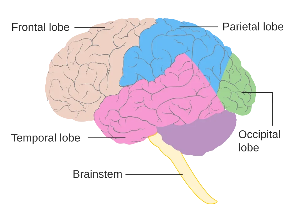
The symptoms depend on the location and growth rate of the tumour. Common symptoms are:
- a first epileptic seizure in adulthood, especially with slow-growing tumours;
- headache that is new, increases over weeks, is worse in the morning or comes with nausea and vomiting;
- gradual loss of function: weakness or loss of sensation in an arm or leg, speech or language problems, loss of part of the visual field;
- changes in thinking, memory, behaviour or personality, often first noticed by those around you.
Warning signs: a first epileptic seizure, rapidly worsening headache with vomiting or drowsiness, sudden loss of function in an arm, leg or speech, or increasing confusion. Go to the emergency department.
How is the diagnosis made?
An MRI scan with contrast shows the location, the size and the probable nature of the tumour. Additional MRI techniques are often used to map the blood supply, the metabolism and the position of important brain pathways and areas. We use these to plan the procedure.
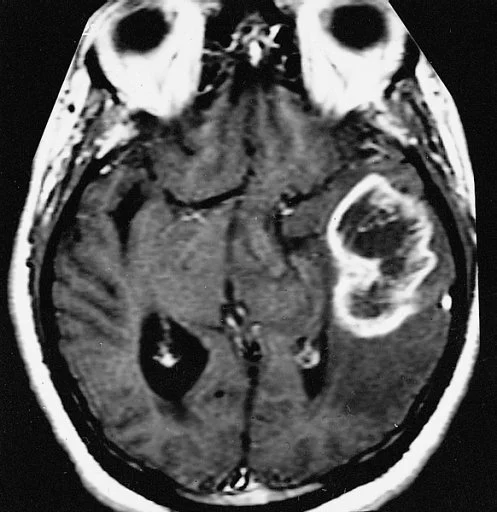
The definitive diagnosis always requires tissue. This is obtained during the operation in which the tumour is removed, or through a brain biopsy (see the separate page) when removal is not safe or not worthwhile. The pathologist examines the tissue under the microscope and tests it for IDH mutation, 1p/19q, MGMT methylation and other features. These molecular tests take a few weeks, but are indispensable: they determine the type, the grade, the prognosis and the choice of further treatment. Every case is discussed at the multidisciplinary oncology meeting.
The operation
Where possible, we try to remove a glioma surgically as completely as possible, without causing new loss of function. This is called maximal safe resection. The more tumour is removed, the better the outlook, for all types; but avoiding permanent loss of function takes priority over the last millimetres of tumour. To achieve this, we use several techniques:
- Neuronavigation: computer guidance during the procedure showing exactly where the tumour and the important brain pathways are located.
- Fluorescence (5-ALA): beforehand you drink a substance that accumulates in high-grade tumour tissue and glows pink under a special light in the microscope. This makes the difference between normal and tumour tissue visible. In the study that proved this, complete removal of the enhancing tumour tissue was achieved in 65% of patients, compared with 36% without fluorescence, with a longer period without regrowth.
- Awake surgery: for tumours in or close to the speech or movement area, part of the procedure can be done while you are awake and carrying out tasks, so that those functions are tested continuously. This is discussed with you in detail beforehand and only makes sense if you feel comfortable with it.
- Intraoperative ultrasound and ultrasonic aspiration: these check how much tumour is still present and remove the tumour tissue precisely, with as little traction as possible on the surrounding tissue.
Even when removal appears complete at first sight, microscopic tumour cells always remain in the surrounding tissue. Sometimes we deliberately leave a small piece of tumour behind, because otherwise we would cause too much damage to important parts of the brain. These tumour remnants are treated with radiotherapy and/or chemotherapy. Within 48 hours of the procedure, an MRI is done to check how much tumour remains.
If the tumour is in a very hard-to-reach place, or has grown too far into areas responsible for, say, speech, we sometimes choose not to operate and to take only a biopsy, in order to preserve quality of life. Radiotherapy and/or chemotherapy then often still give a worthwhile result. Surgery remains our preference, but only if it can be done safely.
For low-grade gliomas (grade 2), we also aim for complete removal if it can be done safely. In some patients, this can halt the disease for years and postpone further treatment. If surgery is too dangerous and the tumour causes few symptoms, we can also monitor with regular MRI scans and intervene if it grows.
Radiotherapy, chemotherapy and new medicines
Further treatment depends on the type, the grade, your age and your condition, and is decided at the multidisciplinary oncology meeting.
Glioblastoma
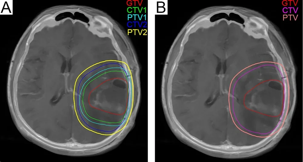
The standard since 2005 is six weeks of radiotherapy combined with the chemotherapy pill temozolomide, followed by six cycles of temozolomide, one per month (the so-called Stupp regimen). In the study that established this standard, median survival rose from 12 to almost 15 months and the number of patients still alive after two years doubled (from 1 in 10 to more than 1 in 4). In patients aged 70 or over, a shorter three-week radiotherapy schedule is used, with or without temozolomide, depending on the MGMT test and their condition. If general health is poor, comfort care may be chosen.
Astrocytoma and oligodendroglioma, IDH-mutant
For a grade 2 tumour that has been completely removed in a patient younger than about 40, further treatment can be postponed and we monitor with MRI. In other cases, and for grade 3, the standard consists of radiotherapy followed by chemotherapy: PCV (three drugs) or temozolomide. In oligodendroglioma, adding PCV to radiotherapy roughly doubles survival. A new development is the medicine vorasidenib, a pill that blocks the mutated IDH enzyme. In a large study of patients with a grade 2 tumour who had not yet received radiotherapy or chemotherapy after surgery, it extended the median time until the tumour grew again from 11 to almost 28 months, and allowed the start of radiotherapy or chemotherapy to be postponed. You can discuss availability and reimbursement in Belgium with the neuro-oncologist.
If the tumour grows back
Almost every glioma grows back sooner or later. We then review all the options again: a second operation (worthwhile in about 1 in 4 patients), further radiotherapy, different chemotherapy (for example lomustine) or taking part in a clinical trial. Supportive medication for epilepsy, swelling (steroids) and fatigue is part of every phase of treatment.
What is the outlook?
This is the hardest question and we answer it honestly, bearing in mind that figures are averages and never an individual prediction.
- Glioblastoma: with the full standard treatment, half of patients live longer than about 15 months; more than 1 in 4 are still alive after two years and about 1 in 10 after five years. Patients with an MGMT-methylated tumour, a younger age, good general condition and complete removal do better than this average.
- Astrocytoma, IDH-mutant, grade 2: in the study that established radiotherapy plus PCV as the standard for higher-risk patients, median survival was more than 13 years, compared with almost 8 years with radiotherapy alone.
- Oligodendroglioma, grade 3: with radiotherapy and PCV, half of patients live longer than about 14 years; in the longest-running studies, more than a third were still alive after twenty years.
- Astrocytoma, grade 3 and 4 (IDH-mutant): in between the two groups above, with survival expressed in years and strongly dependent on additional DNA features.
A cure is rarely possible with a diffuse glioma. The aim of treatment is the longest possible life in the best possible condition, and this is increasingly being achieved.
Recovery and follow-up
After brain surgery you usually stay in hospital for a few days. Fatigue, concentration problems and headache are normal in the first weeks. Physiotherapy, speech therapy or neuropsychological rehabilitation are arranged where needed. After brain surgery and with epilepsy, legal restrictions on driving apply, which we will discuss with you.
Follow-up is done with MRI: every two to three months for a glioblastoma, every three to six months for IDH-mutant tumours, and later less often if the disease remains stable. Shortly after radiotherapy, the scan can temporarily show worsening that is not real growth (pseudoprogression); if in doubt, we repeat the scan after four to eight weeks. Every check-up is discussed at the multidisciplinary oncology meeting, so that we can act quickly if the tumour grows back.
Frequently asked questions
Why don't you just remove everything?
A glioma has no sharp border: tumour cells are also present in brain tissue that looks normal and carries important functions. Removing everything would cause permanent loss of function. We remove as much as can be done safely and treat the rest with radiotherapy and chemotherapy.
What do "grade" and "IDH" mean?
The grade (2 to 4) indicates how fast the tumour grows. IDH is a gene; a mutation in it points to a tumour that grows more slowly and responds better to treatment. A glioblastoma is by definition IDH-wildtype, so without that mutation. Together with the 1p/19q test, both determine the exact type and the treatment.
Is a glioma hereditary?
Almost never. Gliomas arise from random changes in the DNA of brain cells during life. Only in a very small number of families does a hereditary syndrome play a role, and then there are usually other tumours in the family as well.
What is awake surgery, and isn't it frightening?
You are put to sleep for the opening and closing; only during removal of the tumour are you awake and carrying out simple tasks (speaking, naming, moving). Brain tissue itself feels no pain. Most patients find it strange but quite bearable; for those who dread it, we choose a different strategy.
Can I drive after the operation?
Not immediately. After brain surgery and after an epileptic seizure, Belgian law imposes driving bans whose duration depends on the procedure, how long you have been seizure-free and the type of driving licence. We will discuss this with you when you are discharged.
Are there new treatments or trials?
Yes. For IDH-mutant gliomas there is a targeted medicine (vorasidenib); for glioblastoma, trials are under way with immunotherapy, vaccines and new targeted drugs. We discuss taking part in a clinical trial with the neuro-oncologist and, where needed, with a university centre.