
home > treatments > brain haemorrhage and head injury
Brain haemorrhage and head injury
A bleed in or around the brain, or a concussion or contusion
Brain haemorrhages and brain injuries
A brain haemorrhage is an umbrella term. The blood can be in the brain tissue itself, in the space around the brain, or between the membranes covering the brain and the skull. Where it is, why it is there and how much there is together determine whether surgery is needed. Often it is not. This page describes the different types, bleeding after an accident and concussion, and explains when an operation does help.

Which symptoms suggest a brain haemorrhage?
- Sudden loss of function: a paralysed or weak arm or leg, a drooping face, speech problems, reduced consciousness. This is the picture of a stroke; only a CT scan can tell a bleed from an infarct.
- Thunderclap headache: a headache that comes on within seconds and is worse than anything you have ever felt, often with a stiff neck, vomiting or loss of consciousness.
- After a fall or a blow: increasing drowsiness, repeated vomiting, confusion, an epileptic seizure, or someone who drifts off again after a lucid interval.
- Creeping, weeks after a minor fall: headache, slower thinking, an unsteady gait, a weak arm or leg, confusion, especially in older people and in those taking blood thinners.
Warning signs: sudden loss of function in an arm, leg, the face or speech; a headache that comes on within seconds; after a head injury, increasing drowsiness, repeated vomiting, an epileptic seizure or loss of function. Call 112. Do not wait for an appointment.
Bleeding without an accident
Bleeding in the brain tissue (intracerebral haemorrhage)
The most common spontaneous brain haemorrhage arises in the brain tissue itself, usually from a burst small blood vessel in long-standing high blood pressure, sometimes because of blood thinners or a vascular malformation. It causes sudden loss of function: a paralysed arm or leg, speech problems, a drooping face, reduced consciousness. About 10 to 15% of all strokes are such a bleed; the rest are ischaemic strokes (cerebral infarcts) caused by a blocked blood vessel. The CT scan makes the distinction, and that distinction is important because the treatment is the opposite.
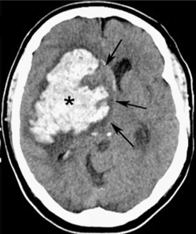
Most of these bleeds are not operated on. The large British STICH trial (more than 1,000 patients) showed that early surgery does not, on average, improve the outcome: after six months, 26% of the patients who had surgery and 24% of those who did not were doing well, and mortality was the same. Brain tissue damaged by the bleed does not recover because we remove the blood. Treatment consists of rapidly lowering the blood pressure, reversing blood thinners and monitoring on a specialised ward.
We do operate when the bleed becomes so large that the pressure inside the skull rises to a life-threatening level, for bleeds in the cerebellum that press on the brainstem (guidelines then recommend immediate removal), and when fluid builds up in the ventricles of the brain and has to be drained with a tube. What is new is that an American trial from 2024 (ENRICH) showed that early removal of a superficial bleed of 30 to 80 ml through a narrow viewing tube, within 24 hours, improves the chance of a good functional recovery after six months. This did not apply to deep bleeds. Whether a patient is eligible for such a procedure is assessed case by case.
Bleeding around the brain (subarachnoid haemorrhage)
A second type is subarachnoid haemorrhage: blood in the space around the brain, usually from a ruptured aneurysm, a bulge in an artery. The typical story is a headache that comes on within seconds and is worse than anything the patient has ever felt. This is always an emergency, because the aneurysm can bleed again. It is secured as quickly as possible, according to current guidelines preferably within 24 hours: through the groin with platinum coils (coiling) or through an opening in the skull with a clip. The choice depends on the shape and location of the aneurysm. For this we refer the patient to UZA or another university centre with a specialised team.
Bleeding after a fall or a blow
In an accident, blood can collect between the skull and the outer membrane of the brain (epidural) or between that membrane and the brain (subdural). An epidural haemorrhage comes from an artery, grows quickly and is a surgical emergency, even if the patient was initially still lucid after the blow. With timely surgery the prognosis is usually good, because the brain itself is mostly not damaged.
An acute subdural haemorrhage usually comes from a vein, is often accompanied by bruising of the underlying brain tissue and therefore has a more serious prognosis, even after a successful operation. Small bleeds in a patient who is awake are followed with repeat scans; large bleeds or bleeds with reduced consciousness are removed urgently through a bone flap in the skull.
Chronic subdural haematoma
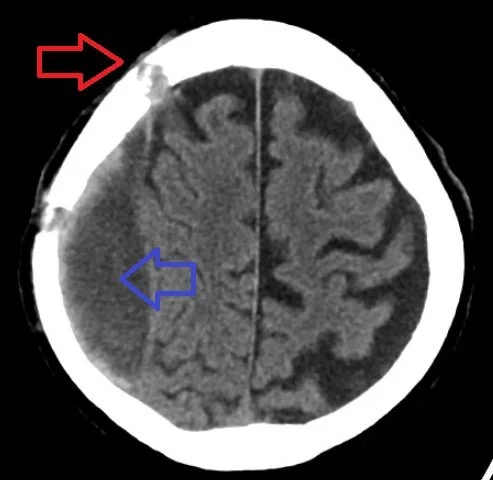
Chronic subdural haematoma is a story of its own and one of the most frequent reasons we operate on older patients. A minor fall, sometimes so slight that nobody remembers it, causes a vein to leak; over weeks the blood collects beneath the membrane of the brain and the body surrounds it with a membrane that itself keeps oozing blood. The symptoms creep in: headache, slower thinking, an unsteady gait, a weak arm or leg, confusion. Blood thinners clearly increase the risk.
The treatment is a small procedure, often under local anaesthesia: through one or two burr holes we let the blood drain out, rinse the cavity and leave a thin drainage tube in place for one to two days. That drain roughly halves the chance that the blood comes back (in the trial that proved this: 9% with a drain versus 24% without). The hospital stay lasts a few days. In about one in ten patients the blood still comes back and a second procedure is needed; in people taking blood thinners, this is slightly higher.
Steroids (dexamethasone) used to be given to prevent this, but a large British trial showed more complications and a poorer recovery; we no longer use them. A new additional treatment is embolisation of the middle meningeal artery: through the groin, the small artery that feeds the bleeding membrane is blocked. Three trials from 2024 showed that this reduces the number of repeat operations after burr-hole drainage (in the largest: 4% versus 11%) and that it slows the growth of small haematomas that are not operated on straight away. It made no difference to the final functional recovery, and the procedure is not yet standard. We discuss it with interventional neuroradiology for patients at high risk of recurrence.
Brain injury without bleeding
Most head injuries are concussions: a temporary disturbance of brain function without any abnormality on the scan. Headache, nausea, difficulty concentrating, irritability and poor sleep are normal in the first days to weeks. In most people these symptoms disappear within a month.
The first two weeks are important. During this period the brain needs time to recover. Allow yourself both mental and physical rest: limit demanding thinking tasks, long screen time, busy surroundings and heavy physical exertion, and avoid alcohol. Enough regular sleep is one of the most important factors for recovery: go to bed at fixed times and plan rest breaks during the day.
Rest does not mean lying in bed or in the dark for weeks, though. For the first 24 to 48 hours, take things as easy as possible; after that, light activity such as walking is good for recovery. Within those first weeks you also build up work, school and screens step by step, as long as the symptoms do not clearly get worse. Whatever makes the symptoms flare up, you do a little less of for a while. Wait until you are completely free of symptoms before taking up contact sports or activities with a risk of falling.
A small proportion of patients have symptoms for longer than four weeks, mainly fatigue and concentration problems. This does not mean there is permanent damage, but it does call for support, sometimes from a rehabilitation physician.
What an operation involves
For a large bleed or severe swelling, we open a flap of bone in the skull under general anaesthesia, remove the blood and stop the bleeding vessels. Usually we put the bone flap back straight away. If the brain is too swollen, we leave the flap out for a few weeks to months (decompressive craniectomy) to give the brain room; it is put back later in a separate operation. After the operation the patient stays in intensive care, where the pressure inside the skull and the blood pressure are closely monitored.
In severe brain injury with pressure that cannot be brought under control, such a decompression saves lives: in the international RESCUEicp trial, 27% of the patients who had surgery had died after six months, compared with 49% without surgery. But some of the additional survivors remain severely dependent on others. We discuss this trade-off, and what the patient would personally find acceptable, with the family before the operation.
How the patient recovers depends more on the damage the bleed or the accident has already done than on the operation itself. Families would rather hear this honestly beforehand than afterwards.
Questions families often ask
Why don't you operate straight away? There is blood in the brain, isn't there?
Because removing blood does not repair the damaged tissue and the operation itself can add damage. We operate when the bleed is pressing or threatens to press on the brain, or when studies show that a particular type of bleed does benefit from surgery, not simply because it is there.
My father is 80 and takes blood thinners. He had a minor fall three weeks ago and is now confused. Could that be related?
Yes, that is the classic story of a chronic subdural haematoma. A CT scan gives a clear answer within an hour. Do not delay.
Should the blood thinners be stopped?
Temporarily, yes, and in an acute bleed their effect is reversed immediately with an antidote. When they can safely be restarted depends on why they were started (an artificial heart valve or a recent thrombosis weighs more heavily than prevention) and is agreed with the cardiologist or GP. After a chronic subdural haematoma this is usually after two to four weeks; after a bleed in the brain tissue in a patient with atrial fibrillation, a longer wait is usual, with guidelines mentioning about seven to eight weeks.
How long does recovery take?
For a concussion, weeks. For a chronic subdural haematoma, often a few weeks to months, and most patients return to how they were before. For a large spontaneous bleed or a severe brain injury, months to more than a year, with rehabilitation, and not always completely. The greatest recovery is seen in the first six months.
Can I drive after a concussion?
Not as long as you have headache, dizziness or concentration problems. After brain surgery or after an epileptic seizure, legal driving restrictions apply, which we discuss with you; the period depends on the procedure and on the type of driving licence.
Can a brain haemorrhage come back?
With a chronic subdural haematoma, in about one in ten. With a bleed caused by high blood pressure, good blood pressure control (target around 130/80) is the best protection against a new one. A treated aneurysm is followed up with imaging; a new bleed from the same aneurysm is rare after that.