
home > treatments > hydrocephalus
Hydrocephalus (water on the brain)
Too much fluid and/or raised pressure in the brain
What is hydrocephalus?
Cerebrospinal fluid circulates constantly in and around the brain. It is produced in the brain's chambers (ventricles), flows around the brain and spinal cord and is then reabsorbed into the bloodstream. If that balance is disturbed, by a blockage in the circulation or impaired absorption, the fluid builds up in the ventricles and the pressure rises. This is called hydrocephalus, popularly known as "water on the brain". It does not only affect children: it also occurs in adults and older people.
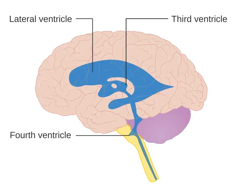
In adults, we roughly distinguish two forms. In obstructive hydrocephalus, a narrowing or a growth is in the way, for example in the narrow channel between the third and fourth ventricles. In normal pressure hydrocephalus (NPH), the form that mainly affects older people, the absorption of the fluid is impaired. The pressure is not clearly raised, but the ventricles slowly expand and press on the surrounding brain pathways. NPH is not rare: in population studies of people over 80, it affects a few per cent, depending on the criteria used.
What symptoms does hydrocephalus cause?
With an acute rise in pressure, for example due to a sudden blockage, headache, nausea and vomiting, drowsiness and blurred or double vision develop. This is a situation that requires prompt treatment.
In older people there is also the gradual form (NPH), with a typical triad of symptoms:
- Walking: increasingly difficult, with small shuffling steps, a wide-based gait and difficulty turning or starting, as if the feet were stuck to the floor. Falls become more common. This is almost always the first and most pronounced symptom.
- Thinking: slower thinking, forgetfulness, less initiative and concentration.
- Bladder: first a strong urge and needing to go often, later loss of control.
By no means everyone has all three symptoms together: in large series, about 6 in 10 patients have the full triad. Because these symptoms are often put down to "just getting older", wear and tear or dementia, NPH is regularly recognised late. That is unfortunate, because it is one of the few treatable causes of this combination.
Warning signs: rapidly worsening headache with vomiting, increasing drowsiness or confusion, double vision, or in someone with a shunt, the sudden return of the same symptoms as before the procedure. This may indicate an acute rise in pressure or a blocked shunt. Go to the emergency department.
How is the diagnosis made?
The first step is brain imaging (CT or MRI). This shows whether the ventricles are enlarged and whether a blockage or growth is the cause. In NPH, the radiologist looks for a typical pattern: wide ventricles combined with narrow fluid spaces at the top of the brain.
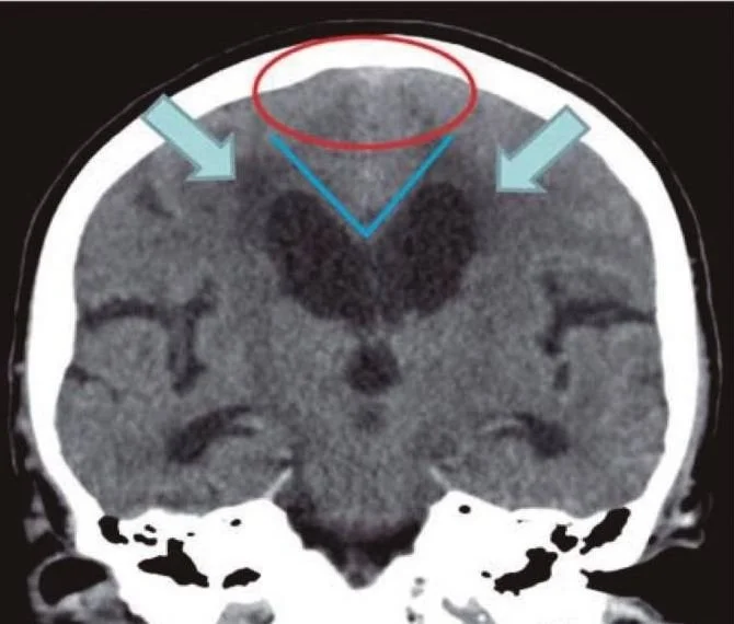
Enlarged ventricles alone are not enough for the diagnosis; they are also often larger with wear and tear and dementia. That is why a trial drainage (tap test) follows: a larger amount of cerebrospinal fluid is drained through a lumbar puncture in the lower back. Walking, and if possible thinking, are measured before and within the following 24 hours, sometimes again in the days after. If you clearly improve, this predicts a good result from a shunt. However, a negative test does not rule out NPH: if there is doubt and a strong suspicion, a lumbar drain can be placed, through which fluid is drained for a few days during a short hospital stay. This tests the effect of drainage more thoroughly.
We also check whether another condition (partly) explains the symptoms, such as Parkinson's disease, a form of dementia, spinal stenosis in the lower back or neck, or problems with the bladder itself. These can exist alongside NPH and help determine how much benefit you can expect from treatment.
How is hydrocephalus treated?
Medication does not work in the long term. If the tests point to a benefit, treatment is surgical. Which procedure is suitable depends on the cause and is discussed with you on the basis of your imaging.
The shunt
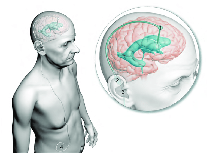
The most commonly used treatment is a ventriculoperitoneal shunt: a thin tube placed in a ventricle through a small burr hole in the skull, connected to a small adjustable valve behind the ear and a second tube that runs under the skin to the abdominal cavity. There the body absorbs the excess fluid. The system lies entirely under the skin and is invisible from the outside. The procedure is performed under general anaesthesia and is relatively short.
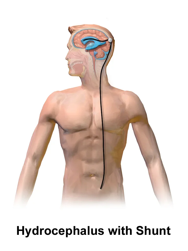
The valve controls how much fluid is drained per day. It can later be adjusted painlessly through the skin with a magnetic adjustment device, without a new procedure. This allows us to match the drainage to your symptoms and correct over- or underdrainage. The system also contains a reservoir that lets us test whether the shunt is still working properly.
The evidence for shunting in NPH was strengthened in 2025 by a double-blind study of 99 patients: everyone received a shunt, but in half of them the valve was set to "closed" for the first three months. In the group with a working shunt, walking speed improved clearly, in about 8 in 10 patients; in the placebo group in about 1 in 4. Patients with a working shunt also fell less often.
Endoscopic third ventriculostomy
In obstructive hydrocephalus, where a narrowing or growth blocks the circulation, an endoscopic procedure can make a shunt unnecessary. Through a small burr hole, an opening is made with a camera in the floor of the third ventricle, so that the fluid finds a new route. In series of patients with a narrowing of the channel (aqueductal stenosis) or a growth, this succeeds in about 8 to 9 in 10. The big advantage: no implant that can become blocked or infected. In NPH this technique works less well and the shunt is the standard.
What is recovery like?
The hospital stay usually lasts a few days. You can get out of bed soon; the small wounds on the head and abdomen heal in one to two weeks. In NPH, walking often improves first, sometimes within days. Memory and continence follow more slowly and not always completely. Across all studies, about 7 in 10 patients improve; for walking this is about 7 in 10, for thinking and bladder control about half. The earlier the treatment and the fewer other conditions play a role, the greater the benefit. Physiotherapy after the procedure helps to build up walking again.
The first check-up takes place a few weeks after the procedure, with a CT scan to assess the position of the catheter and the size of the ventricles. The valve is then adjusted if necessary. A shunt requires lifelong follow-up: with new or recurring symptoms, a shunt problem must always be considered.
What are the risks?
Besides the general risks of an operation under anaesthesia, the main shunt-related problems are as follows, with figures from a review of 54 studies involving more than 4,000 NPH patients:
- Blockage or malfunction of the shunt (about 6 in 100), with return of symptoms. The shunt is then checked and replaced if necessary.
- Overdrainage: too much fluid is drained. This causes headache on standing up, and can lead to a collection of fluid or bleeding under the membrane covering the brain (subdural haematoma), in about 7 in 100. This can usually be corrected by adjusting the valve; sometimes a small procedure is needed.
- Infection of the shunt (about 2 in 100), which requires removal of the system and treatment with antibiotics, followed by a new shunt.
- Repeat operation for one of the reasons above: in the same studies, in about 12 in 100 patients with a ventriculoperitoneal shunt.
Overall, about 1 in 5 patients has a complication, usually mild and reversible. Death due to the procedure is very rare (about 2 in 1,000). With a third ventriculostomy the complication rate is lower, but the opening can close up again. You and those close to you will receive clear instructions about which signs need attention.
Frequently asked questions
Is a shunt visible?
No. The entire system lies under the skin. In slim people, the valve behind the ear can sometimes be felt as a small bump.
Does the shunt last a lifetime?
Often, yes. If there are problems, it can be repaired or replaced. Regular follow-up is part of it, and with recurring symptoms the shunt must always be checked.
My elderly relative is walking worse and worse and becoming forgetful. Could this be NPH?
That is exactly the combination in which NPH should be considered, especially if walking is the first and main symptom. A scan and a trial drainage give a clear answer, and unlike many other causes, this condition is treatable.
What if the trial drainage gives no improvement?
A negative test does not rule out NPH. If suspicion is strong, a lumbar drain for a few days can give more certainty. If that is also negative, the chance of benefit from a shunt is small and we look further for another explanation of the symptoms.
Can I have an MRI scan with a shunt?
Yes, with today's adjustable valves that is possible. Always mention that you have a shunt: after an MRI, the valve setting is checked and reset if necessary, because a strong magnetic field can change it.
Do you recognise these symptoms in yourself or someone close to you?
Discuss it with your GP or book an appointment for further evaluation. Bring any available imaging with you.
Related treatments
All treatments →