
home > treatments > cervical (neck) fusion
Cervical fusion
Fixing the neck vertebrae
What is a cervical fusion?
In a cervical fusion, two or more neck vertebrae are fixed to each other, so that they grow together into one stable unit in the months after the procedure. This is usually done together with freeing a trapped nerve or the spinal cord. A fusion can be done in two ways:
- From the front (anterior): the most commonly performed procedure is anterior cervical discectomy and fusion, abbreviated to ACDF. The surgeon works through an incision of about four centimetres in a skin crease at the front of the neck, removes the intervertebral disc and places a cage in its place.
- From the back (posterior): the vertebrae are fixed with screws and rods through an incision in the middle of the back of the neck. This is mainly considered for narrowing over several levels, instability, after an accident or when the neck needs extra support after an extensive decompression.
Sometimes both are combined. Which route fits best depends on where the pressure is, how many levels are involved and the alignment and stability of your neck.
When do we suggest this operation?
The procedure is usually suggested to free a trapped nerve root. A protruding disc or bony ridge then presses on a nerve running to the arm. This causes pain radiating to the shoulder, arm or hand, sometimes with tingling or weakness. In most people these symptoms disappear within a few months with painkillers, physiotherapy and sometimes an injection. We only consider an operation if the pain has not improved after six to twelve weeks, or earlier if the strength in the arm or hand decreases.
The second reason is pressure on the spinal cord itself, myelopathy. This causes different symptoms: clumsy hands, difficulty with buttons or writing, an unsteady gait and sometimes bladder problems. A spinal cord that has been under pressure for a long time recovers poorly. In moderate or severe myelopathy, an operation is recommended and we wait less long.
A fusion from the back is mainly considered for:
- narrowing of the canal over three or more levels, or pressure coming mainly from behind;
- instability or slippage between vertebrae;
- fractures and injuries after an accident;
- a neck that might otherwise sag forward after an extensive decompression, or to stop further narrowing caused by an overly mobile, worn segment.
Neck pain without radiating pain and without spinal cord symptoms is rarely a reason for fusion. The result is then unpredictable and our advice is usually not to go ahead with it.
What does the procedure involve?
Fusion from the front (ACDF)
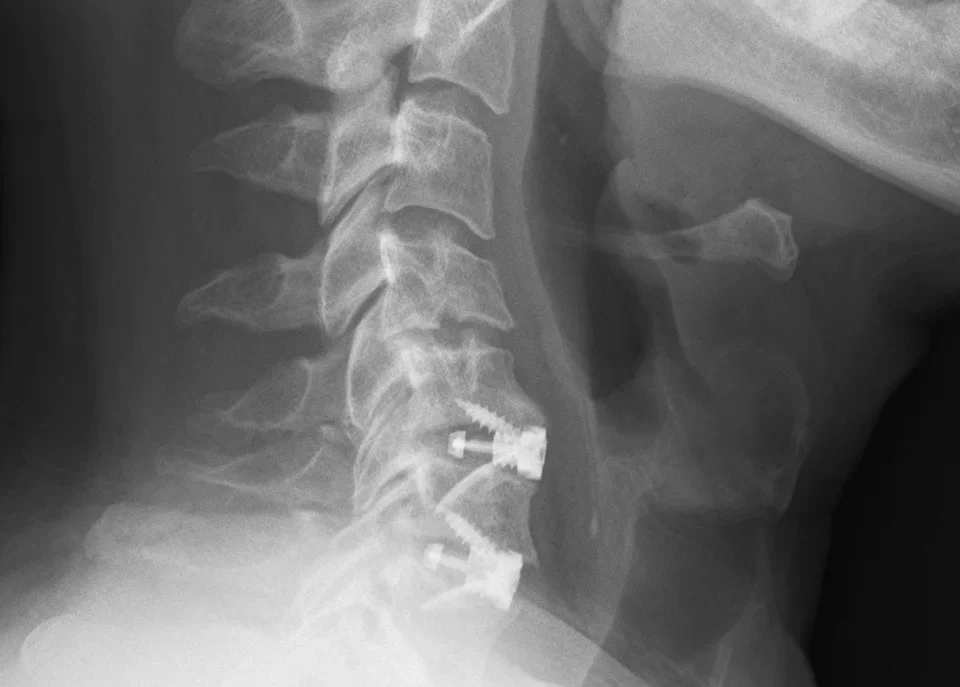
The procedure is performed under general anaesthesia. Through the incision at the front of the neck, the windpipe and gullet are gently moved to one side and the carotid artery to the other. This is how the surgeon reaches the front of the spine. The intervertebral disc is removed completely under the operating microscope, until the nerve and spinal cord are free. A cage made of synthetic material or titanium and filled with bone substitute is placed in the space created. The cage keeps the vertebrae apart and allows them to grow together over the following months. Sometimes a small plate is screwed over it for extra strength.

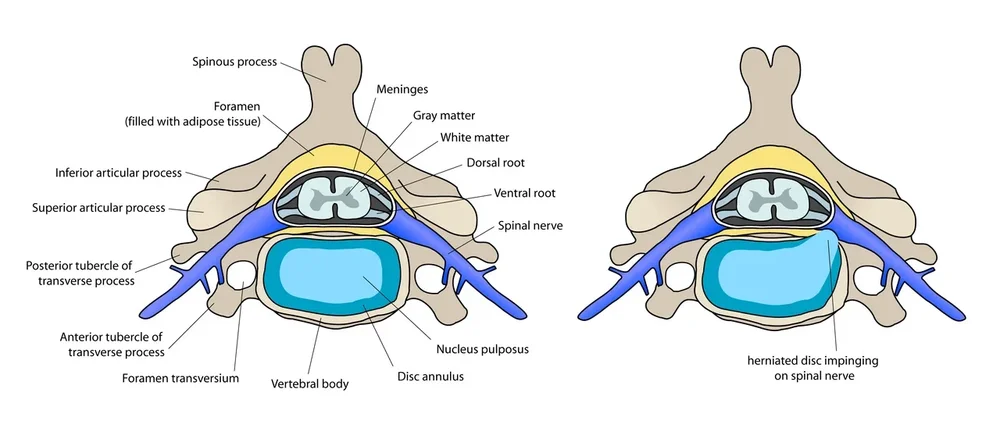

One level takes about one to one and a half hours. Most patients can go home the day after the procedure, sometimes only on the second day. A neck collar is usually not needed. You can read more about the disc herniation itself on the page about cervical disc herniation.
Fusion from the back (posterior fixation)
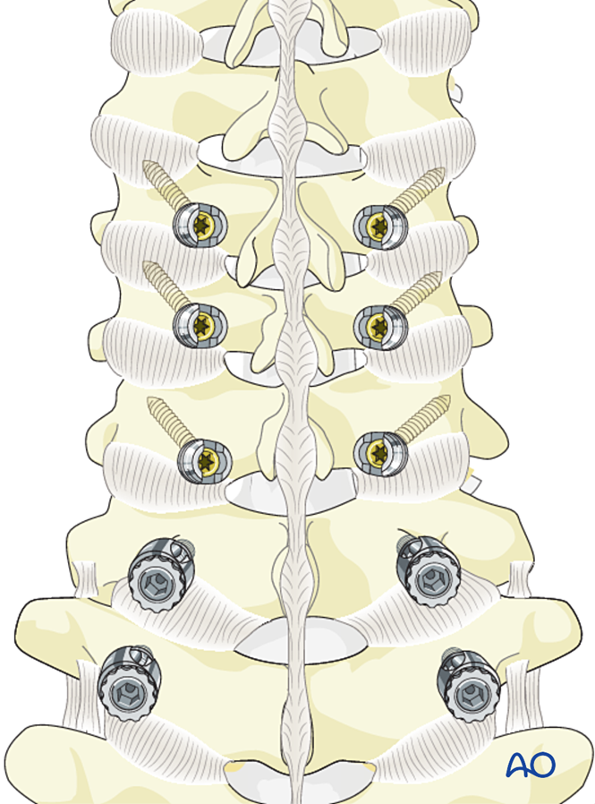
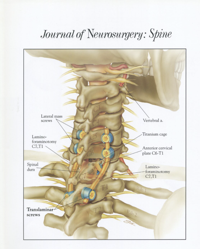
You lie on your front, with your head firmly supported. Through an incision in the middle of the back of the neck, the neck muscles are moved aside down to the vertebral arches. Where necessary, the spinal cord or nerve root is freed first. Screws are then placed in the vertebrae, using navigation, and connected on both sides with a rod. Bone material is applied over the bone so that the vertebrae grow together over the following months. Depending on the number of levels, the procedure takes two to four hours; the hospital stay is usually somewhat longer than after a procedure from the front.

Alternative: procedure from the back without fusion
For a disc herniation or bony ridge at the side that traps a single nerve root, the nerve can also be freed from the back (foraminotomy), without removing the disc and without fusion. A recent Dutch study showed that this technique gives results at least as good as ACDF after one year. With pressure on the spinal cord or a centrally located disc herniation, this is not an option. For narrowing over several levels, a laminoplasty is sometimes an alternative to a fusion from the back; you can read more about this on the page about cervical spinal stenosis.
What can you expect after the operation?
In most patients the arm pain is greatly reduced straight after the procedure. Tingling and weakness recover more slowly, over weeks to months, and not always completely after long-standing compression. Spinal cord symptoms improve more slowly; there, the first aim is to prevent further deterioration.
After a procedure from the front, swallowing is somewhat more difficult for the first few days and the voice may sound hoarse, because the gullet and the nerve to the vocal cords lie close to the operating area. In almost everyone this disappears within a few weeks. After a procedure from the back, it is mainly the wound and the neck muscles that are painful in the first weeks; recovery there usually takes somewhat longer.
- Desk work: can usually be resumed after two to four weeks after a procedure from the front; often somewhat later after a fusion from the back.
- Physical work: usually after six to twelve weeks.
- Driving: as soon as you can turn your neck without pain and no longer need strong painkillers, usually after two weeks.
- Bone healing: the bone fuses in three to six months. Until then, avoid heavy loads and contact sports.
- Follow-up: a consultation with an X-ray after six weeks and after three months.
What are the risks?
After a procedure from the front, temporary swallowing problems and hoarseness are the most common. In large reviews of studies, swallowing difficulties are reported as a complication in about 1 in 10 patients and hoarseness in about 2 in 100; in the first few days a much larger proportion are mildly affected. Less common are:
- bleeding in the neck after the operation (about 1 in 100), which sometimes requires a second procedure;
- a wound infection (less than 1 in 100);
- a leak of cerebrospinal fluid through a small tear in the membrane around the nerves (less than 1 in 100);
- permanent hoarseness due to injury of the vocal cord nerve (about 1 to 3 in 100);
- an injury to the gullet: very rare (less than 1 in 100), but serious.
After a fusion from the back, the emphasis is different:
- wound problems and infection are more common than after a procedure from the front;
- temporary shoulder weakness (C5 palsy) in about 5 to 8 in 100 patients, usually recovering within a few months;
- neck pain and stiffness for the first weeks to months;
- rarely, an injury to a blood vessel or nerve from a screw; navigation reduces this risk.
A spinal cord injury with paralysis is very rare, but not impossible. We discuss this openly beforehand.
In some patients the vertebrae do not fuse. This is called pseudarthrosis. After an ACDF, across all studies, this concerns about 1 in 10, more often in fusions over several levels and in smokers. It does not always cause symptoms, but may make another procedure necessary.
In the long term, the levels above and below the fusion can wear more quickly, because they take over the movement. In a long-term study, about 1 in 4 patients developed new symptoms at an adjacent level within ten years (about 3 in 100 per year). Most patients are never troubled by this; a minority need a second procedure years later.
Questions patients often ask
Will my neck become stiffer?
After one fixed level, less than you might expect: the other levels largely make up for it. With a fusion over two or three levels, and certainly after a fusion from the back over several levels, you will notice it, especially when looking over your shoulder.
How big is the scar?
From the front about four centimetres, in a natural skin crease at the front of the neck; after a year it is barely visible in most people. From the back the incision is longer, depending on the number of levels, but it is less noticeable on the back of the neck.
Will the implants ever need to be removed?
No. The cage and screws stay in place for life. They do not cause problems with metal detectors at the airport, and an MRI scan can go ahead as normal.
Can I try an injection first?
Yes, as long as there is no weakness and no myelopathy. An injection around the nerve root relieves the pain in some patients, sometimes enough to avoid an operation. It does not change the compression itself.
Why from the front and not from the back?
With a disc herniation or bony ridge at one or two levels, the disc and bony ridges trapping the nerve lie at the front. From the front they can be reached directly, without going through the neck muscles, which causes less pain after the procedure. The back is better suited for narrowing over several levels, instability and after accidents, and for a foraminotomy for narrowing at the side.
What if I do nothing?
With arm pain without weakness, that is often not a problem. The symptoms may disappear, persist or come back. With myelopathy, waiting is riskier, because loss of spinal cord function often no longer recovers fully. The same applies to severe weakness due to marked pressure on a nerve root.