
home > treatments > cervical (neck) stenosis
Cervical stenosis
Narrowed canal in the neck
What is cervical spinal stenosis?
Wear and tear of the spine, which happens to everyone with age, can narrow the canal through which the spinal cord runs in the neck. This is called cervical spinal stenosis.
The narrowing develops because intervertebral discs bulge or calcify, facet joints and ligaments thicken, and bony outgrowths form on the vertebral bodies. Sometimes a canal that is narrow from birth plays a part, so that symptoms appear at a younger age. In some people the alignment of the neck also changes, which increases the pressure on the spinal cord.
The big difference from the lower back is that the neck contains the spinal cord. Pressure on the spinal cord, called cervical myelopathy, is more serious than pressure on a single nerve root, because the spinal cord carries all signals to the arms, legs and bladder. Moreover, a damaged spinal cord recovers only to a limited extent.

What symptoms does cervical stenosis cause?
The symptoms usually develop gradually, over months to years, and are therefore often put down to getting older. Typical symptoms are:
- tingling, a numb or swollen feeling in the hands, often on both sides;
- reduced fine motor skills: doing up buttons, writing, picking up small objects, putting a key in a lock;
- unsteady or stiff walking, a feeling of walking "on cotton wool", difficulty on stairs or in the dark, stumbling more often;
- weakness in the arms or legs;
- pain radiating from the neck to the shoulder or arm, when a nerve root is also trapped;
- at a later stage: difficulty holding urine or, conversely, passing urine.
Neck pain is not a requirement. Many patients with a clear narrowing have little or no pain, but do have the functional symptoms listed above.
Warning signs: rapidly worsening symptoms, sudden weakness in the arms or legs, or new bladder problems are a reason to contact your doctor or the practice promptly. A fall or a sudden neck movement followed by worsening symptoms also requires prompt assessment.
How is the diagnosis made?
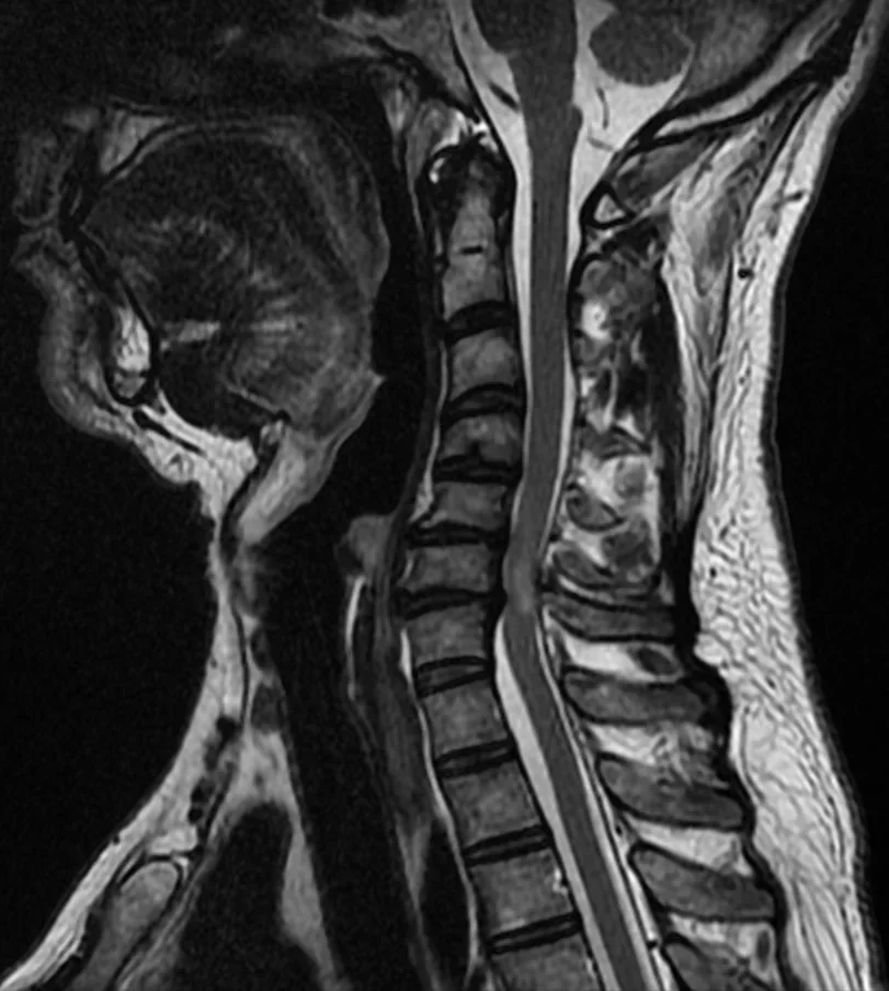
The physical examination focuses on reflexes, strength, sensation and gait. An MRI of the neck is the most important scan: it shows the narrowing and whether the spinal cord itself already shows signs of damage. X-rays in forward and backward bending, and sometimes a CT scan, are often also requested to assess the alignment and stability of the neck.
A nerve conduction study (EMG and/or MEP/SEP) is often done as well. It measures how well signals travel through the spinal cord and nerves and helps rule out other causes, such as carpal tunnel syndrome, polyneuropathy (for example in diabetes) or another neurological condition. For this we consult with colleagues in neurology and physical medicine.
We express the severity of the myelopathy with a score (mJOA) that measures hand function, gait, sensation and bladder function. This score helps determine our advice.
Do I need surgery?
Not every narrowing requires an operation. International guidelines (AO Spine and the Cervical Spine Research Society, 2017) make the following distinction:
- Narrowing without spinal cord symptoms: preventive surgery is not recommended. Follow-up is needed, however, because about 8 in 100 of these patients develop myelopathy within a year, and about 23 in 100 within a period of four years. Those who also have radiating arm pain are at higher risk; in that case surgery may be considered.
- Mild myelopathy: both an operation and a supervised rehabilitation programme with close follow-up are reasonable choices. If you deteriorate under non-surgical treatment, an operation is advised.
- Moderate or severe myelopathy: an operation is recommended, even if the symptoms still seem manageable to you.
The reason is the natural course: without treatment, 2 to 6 in 10 patients with myelopathy deteriorate further within 3 to 6 years, sometimes gradually, sometimes in steps after a minor fall or a sudden movement. Lost function often does not return. Physiotherapy, medication or injections can relieve pain, but do not remove the pressure on the spinal cord.
Which operations are possible?
The principle is always the same: giving the spinal cord and nerve roots more room. The choice depends on where the pressure is (in front of or behind the spinal cord), the number of levels, the alignment and stability of the neck, and your general health. In a large North American study of 163 patients, an operation from the front and an operation from the back gave comparable recovery after one and two years; the pattern of complications did differ, however (more temporary swallowing problems with the front approach, more wound problems and temporary shoulder weakness with the back approach). All the techniques below are offered in the practice; we choose the most suitable one together with you.
Decompression from the front (ACDF or corpectomy)
Through a small incision in the neck, the intervertebral disc (ACDF) or part of the vertebral body (corpectomy) is removed. The space is filled with a small cage or a bone graft and the vertebrae grow together (fusion). This is the preferred technique for pressure at the front limited to one or two levels, or for a neck that is tilted forward.
Decompression from the back
With extensive narrowing or narrowing that is mainly at the back, the pressure is relieved from behind. Under the microscope, only the structures causing the pressure are removed (thickened ligaments, bony outgrowths), while stability is preserved.
Decompression with fixation
Sometimes a fusion is needed, in which the decompression is combined with spinal screws and rods and the vertebrae grow together. This is the case when the neck needs stability: with slippage between vertebrae, a neck tilted forward (kyphosis), marked wear of the facet joints or after an accident. A fusion may also be needed to stop further narrowing: if a worn, overly mobile segment keeps pressing against the spinal cord with every movement, fixing that segment removes this irritation and prevents the neck from sagging further after the decompression.
Laminoplasty with preservation of the midline
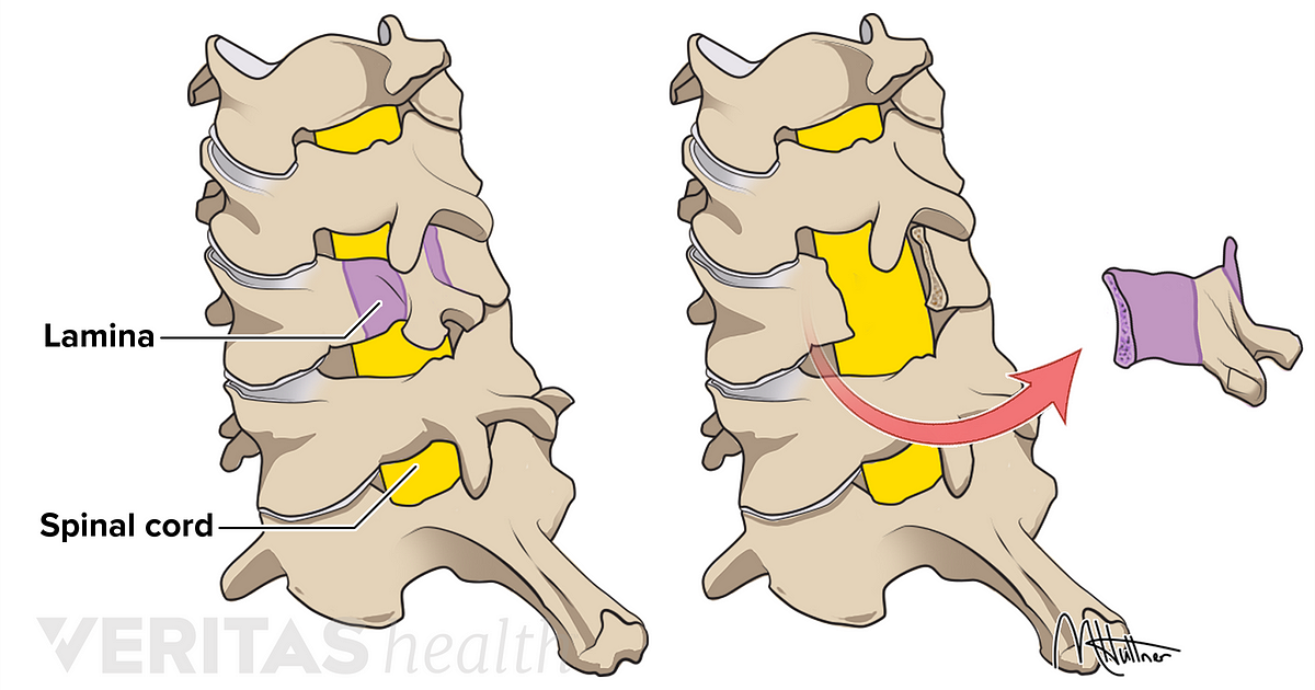
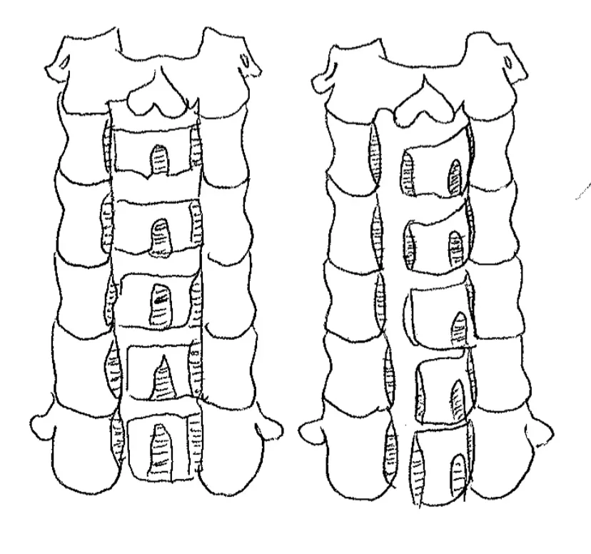
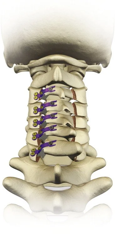
Dr Lavrysen specialises in laminoplasty with full preservation of the midline structures. In a laminoplasty, the vertebral arches are not removed but opened on one side like a door, while the other side acts as a hinge. Small titanium plates hold the door open, so that the canal is permanently widened. The spinous process, the ligaments between them and the neck muscles attached to them remain intact; they are not detached or cut. Often no screws are needed in the vertebrae.
What does the research say? In comparative studies, spinal cord function recovers as well after a laminoplasty as after a laminectomy with or without fixation, with less temporary shoulder weakness (C5 palsy) and fewer superficial wound infections. In the large North American study mentioned above, laminoplasty was associated with fewer complications than laminectomy with fixation; that comparison was not randomised. When the midline structures and muscle attachments are preserved, several studies show less neck pain after the operation and better preservation of neck mobility. That evidence comes mainly from smaller, non-randomised studies (the only randomised study found no difference in neck pain after one year), but no disadvantages were found as long as the spinal cord is adequately decompressed. The technique is suitable when the neck is stable and has a normal or straight alignment; with a markedly forward-tilted neck (kyphosis) or pressure that is mainly at the front, a different approach is better.
What can you expect from the operation?
The first aim is to prevent further deterioration and stabilise the current situation. In many patients something also improves: the tingling, hand function or walking. How much depends on how long and how severely the spinal cord has been under pressure and on what the MRI shows. A full recovery is not guaranteed, and symptoms that have been present for years rarely disappear completely.
The spinal cord recovers slowly; improvement can continue for up to a year after the operation. Radiating arm pain usually improves fastest, sensory disturbances and walking problems slowest.
What are the risks?
The general risks of an operation are infection, post-operative bleeding and wound problems. Specific to these procedures:
- Temporary shoulder weakness (C5 palsy) after a decompression from the back: in about 5 to 8 in 100 patients (from the front about 3 in 100). In most cases this recovers within a few months; permanent weakness affects about 1 in 100.
- Leakage of cerebrospinal fluid through a small tear in the dural sac, usually repaired during the operation.
- Temporary hoarseness or swallowing problems after an operation from the front: swallowing problems are common in the first weeks and resolve in almost everyone; permanent hoarseness due to injury of the vocal cord nerve is rare (about 1 to 3 in 100).
- Failure to fuse or loosening of implants after fixation or fusion, which sometimes requires a second operation.
- Deterioration of spinal cord function: very rare, but not impossible.
We discuss these risks during the consultation and weigh them against the risk of not operating.
Practical matters: before and after the operation
- Hospital stay: under general anaesthesia; usually a few days, depending on the technique and your recovery.
- Movement: you may stand up and walk from the first day; moving early helps recovery.
- Neck collar: with most techniques not needed, or only briefly; after a fixation a soft collar may be advised for a few weeks.
- Wound: after a few days you may take a short shower; no bath or swimming pool until the wound has fully closed.
- Driving: only when you can turn your neck easily and no longer need strong painkillers, usually after a few weeks.
- Work: desk work usually after a few weeks; heavy physical work only after a few months and in consultation.
- Physiotherapy: after the wound has healed, focusing on posture and neck mobility and, if needed, on walking and hand training.
- Follow-up: a first check-up a few weeks after the procedure, with imaging if needed.
Get in touch if you have a fever, increasing redness or fluid leaking from the wound, suddenly increasing weakness or sensory disturbances, or sudden bladder problems.
Frequently asked questions
Am I too old for this operation?
Age alone is rarely an obstacle; your general health is what counts. Because myelopathy continues to worsen without treatment, an operation is often the best choice even at an older age.
Will I be completely back to my old self after the operation?
That depends on the damage the spinal cord has already sustained. The operation stops the deterioration; any recovery beyond that varies from person to person. The earlier the procedure, the greater the chance of recovery.
Will the operation help my neck pain?
Mainly for radiating arm pain and spinal cord symptoms. For neck pain caused by wear and tear, the effect is smaller.
Will I still be able to move my neck after the operation?
After a decompression or laminoplasty, mobility is largely preserved. After a fusion or fixation you lose mobility at the fixed levels; the other levels usually take over most of this.
What if I do not want an operation?
That is your choice. We will then discuss honestly what you can expect and follow you up closely, so that we can intervene in time if the symptoms get worse.
What if the spinal cord already looks damaged on the MRI?
Even then an operation is usually worthwhile, because it prevents further damage. Some of the existing damage may remain; the MRI helps us estimate how much recovery you can expect.