
home > treatments > lumbar disc herniation
Slipped disc in the lower back
Lumbar disc herniation
What is a lumbar disc herniation?
Between the vertebrae of your back are the intervertebral discs: shock absorbers with a tough fibrous ring and a soft core. Through wear and tear or overload, this ring can tear, allowing part of the core to bulge outwards. This is called a disc herniation (or slipped disc). If this fragment presses on a nerve root, the typical symptoms appear.
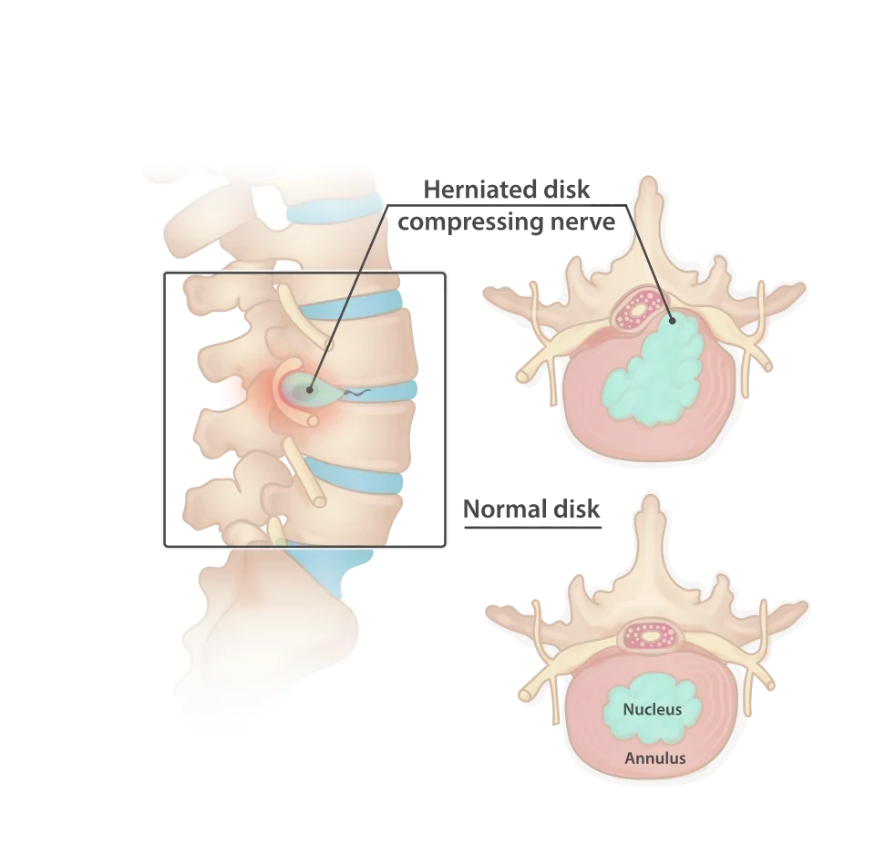
What symptoms does a lumbar disc herniation cause?
- Pain from the back or buttock that radiates into the leg, sometimes down to the foot ("sciatica")
- Tingling or numbness in the leg or foot
- In more severe cases: loss of strength, for example a foot drop
Warning signs: loss of bladder or bowel control, or numbness in the saddle area (between the legs and around the buttocks). In that case, contact a doctor or an emergency department immediately.
Does a disc herniation need surgery?
Usually not. Most disc herniations heal on their own within six to twelve weeks. Initial treatment consists of pain relief, anti-inflammatories and staying active as far as the pain allows, with close follow-up by your doctor. Surgery is only proposed when symptoms persist despite this treatment, or sooner if weakness is increasing.
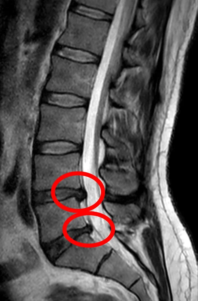
Important to know beforehand: the operation is mainly effective against leg pain. It has less effect on back pain itself. Dr Lavrysen discusses this with you openly, so that you can decide with realistic expectations.
How is the operation performed?
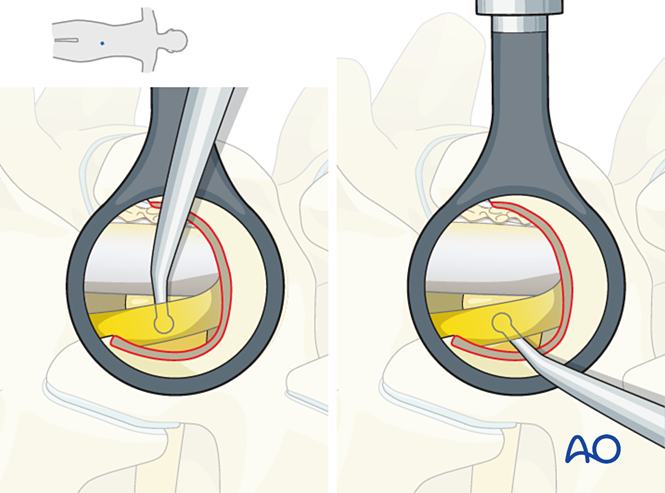
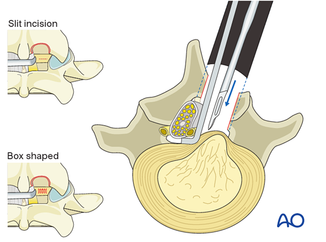
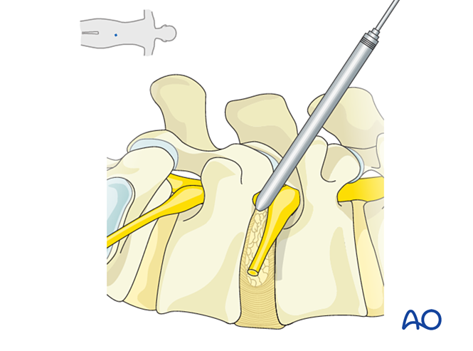
The standard technique is the microsurgical discectomy (microdiscectomy): through a small incision in the back, the muscles are not cut but gently moved aside, after which the trapped nerve root is freed under the operating microscope and the herniation removed. In selected cases the operation can be performed endoscopically, through a working channel of less than one centimetre.
How quickly will I recover?
A few hours after the operation you are back on your feet. Walking is encouraged from day one; office work and driving are usually possible after about two weeks, physical work and sport after around six weeks, always in consultation.
What are the risks?
As with any operation: post-operative bleeding, infection, a small tear in the dura (the membrane surrounding the nerves), and rarely nerve damage. In addition, in 5 to 10% of patients a new herniation may occur at the same level over time. All risks are discussed with you before the operation.
Frequently asked questions
How long does the operation take?
On average about one hour.
Will I have a microsurgical or an endoscopic operation?
That is determined by the MRI: the position and type of the herniation. Both are minimally invasive; Dr Lavrysen will explain the choice during the consultation.
Will my back pain go away too?
The operation targets the leg pain. Back pain due to wear and tear improves less predictably. Dr Lavrysen will discuss the further options with you; these are usually non-surgical.