
home > treatments > lumbar (lower back) stenosis
Lumbar spinal stenosis
Narrowed canal in the lower back
What is spinal stenosis?
Wear and tear of the spine, which happens to everyone to some degree with age, can narrow the canal through which the nerves run. This is called spinal stenosis (narrowing of the spinal canal).
The narrowing develops because ligaments, facet joints, intervertebral discs and vertebral bodies thicken and change shape, or sometimes position. As a result, the spinal canal, the space for the spinal cord and nerves, gradually becomes smaller. The nerves come under pressure, and that causes symptoms.
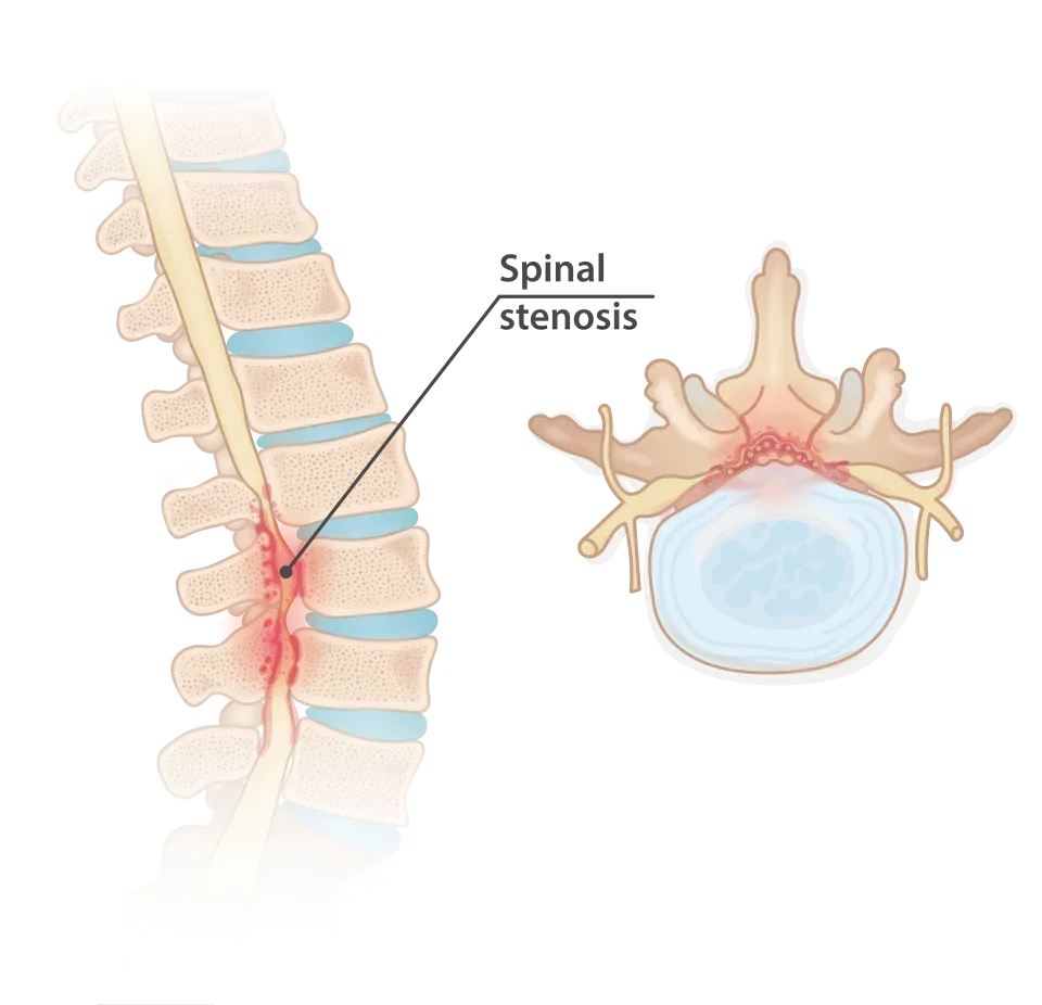
The symptoms differ depending on whether the narrowing is in the neck or in the lower back. This page is about the lower back; for the neck, please see the page on cervical spinal stenosis.
What symptoms does stenosis of the lower back cause?
The canal in the lower back no longer contains spinal cord: the cord ends at the level of the junction between the thoracic and lumbar vertebrae. Nerve roots to both legs do run through it, however. A narrowing therefore typically causes:
- pain, heaviness or cramps in both legs (sometimes one leg) that come on or get worse when walking or standing for a long time;
- an ever shorter distance you can walk without leg symptoms;
- sometimes numbness or loss of strength in the legs;
- relief when standing still, sitting or bending forward. Bending over, walking bent forward (for example behind a shopping trolley) or cycling are often easier than walking.
This pattern is called neurogenic claudication. Back pain may be present, but it is not the characteristic symptom.
Warning signs: sudden loss of bladder or bowel control, numbness in the genital area or around the anus, or rapidly increasing weakness in the legs require urgent assessment. In that case, go to the emergency department.
How is the diagnosis made?
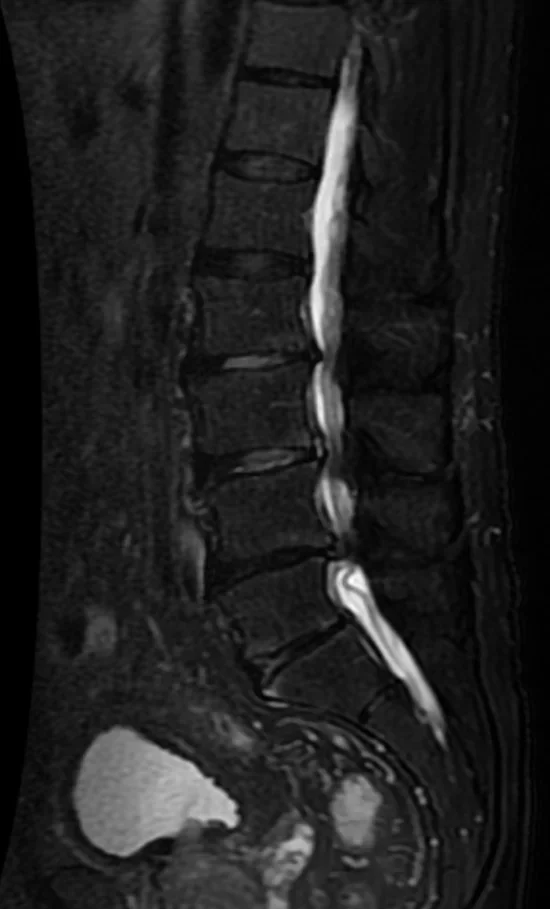
We examine you and review any imaging of the spine that is already available, or request it. An MRI is the most suitable scan to confirm the narrowing and assess its severity. An additional nerve conduction study (EMG or MEP/SEP) is often arranged to refine the diagnosis.
It is important to rule out other causes of leg symptoms when walking, such as narrowed arteries in the legs (vascular claudication), hip osteoarthritis or conditions of the nerves themselves (polyneuropathy). For this we consult with colleagues in vascular surgery, neurology and physical medicine.
Does stenosis need surgery?
Not always. A narrowing on the MRI is not in itself a reason to operate; your symptoms and the limitations in your daily life are. With mild to moderate symptoms, the course without surgery remains favourable in about a third to half of patients, and rapid or severe neurological deterioration is rare. So there is time to try non-surgical treatment first: appropriate exercise and physiotherapy, pain relief and adapting your activities. Epidural injections are not recommended for neurogenic claudication caused by central narrowing; for radiating pain from a single nerve root, however, they can help temporarily.
Surgery is considered for severe, symptomatic narrowing in which walking distance is severely limited or weakness develops, and when non-surgical treatment does not help enough. In comparative studies, patients who had surgery had clearly less pain and fewer limitations in the first years than patients who did not; in the longer term (from 5 to 8 years onwards) the differences become smaller, partly because many patients who did not have surgery end up having it after all. We make the decision together, based on your symptoms, your general health and your expectations.
What does the operation involve?
For severe, symptomatic narrowing in the lower back, a decompression is performed. Traditionally, the entire back part of the vertebral arch is removed (laminectomy).
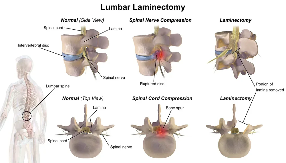
Dr Lavrysen specialises in microscopic decompression with preservation of the midline. Through a small approach on one side, and under the operating microscope, only the structures pressing on the nerves, such as bony outgrowths and thickened ligaments, are removed, on both sides if necessary. The structures needed for the stability of the spine (spinous process, midline ligaments, a large part of the facet joints) are preserved. In this way the dural sac, the sac containing the nerves, and the nerve roots themselves are freed.
In comparative studies, this approach gives as good an improvement in leg symptoms and daily functioning as the traditional laminectomy, with less chance of the spine becoming unstable after the procedure. Some studies also show slightly less back pain after the operation, but that difference is small.
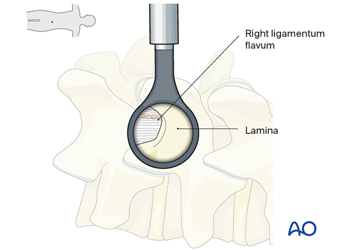
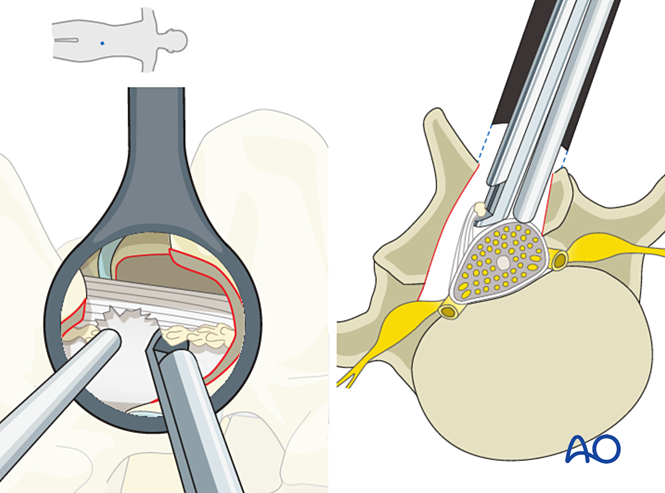
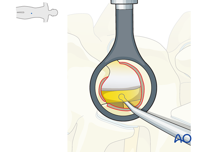
In most cases screws are not needed, because the stability of the vertebrae is preserved. Large studies show that adding a fusion (screws and rods) to the decompression in stenosis without clear instability does not give a better result, but does mean a longer operation and a longer hospital stay. Even with a limited slippage of a vertebra (degenerative spondylolisthesis), a decompression with preservation of the midline is often sufficient; a fusion is reserved for patients with clear instability or a marked slippage. You can read more about this on the page about lumbar fusion.
The opening that is created is not filled with artificial material. After the wound is closed, the muscles and scar tissue protect the nerves; that tissue is strong enough.
The aim of the operation is to reduce the leg symptoms and increase walking distance. Back pain from general wear and tear often does not disappear with this procedure; it is usually treated in a different way.
What are the risks?
Decompression is a safe procedure, also at an older age, but it is not without risks. In studies, a complication is reported in about 1 to 2 in 10 patients who have the operation, usually mild and temporary.
- Small tear in the membrane around the nerves (dural tear) with leakage of cerebrospinal fluid; this is usually repaired during the procedure and can cause temporary headache.
- Post-operative bleeding or wound infection.
- Nerve injury with weakness or altered sensation: rare.
- Insufficient improvement or return of symptoms, for example due to new narrowing at the same or another level. In long-term studies, about 1 in 5 patients needed another back operation within a few years.
- General risks of an operation under general anaesthesia, depending on your age and health.
What is the recovery like?
You may stand up and walk on the day of the procedure or the day after. Short walks are possible from the first days and are encouraged; the distance is built up gradually. The hospital stay is usually short. For the first weeks, avoid heavy lifting and sudden twisting. Desk work and driving can usually be resumed after a few weeks, heavier work later, in consultation. Physiotherapy can support your recovery. Leg symptoms often improve quickly; numbness or weakness recovers more slowly and not always completely.
Frequently asked questions
Am I too old for this operation?
Age alone is rarely an obstacle; your general health is what matters. The minimally invasive approach makes the procedure very feasible even at an older age.
Will the operation also help my back pain?
The operation mainly helps with the leg symptoms and the limited walking distance. The effect on back pain caused by wear and tear is smaller.
How soon can I walk again?
Almost immediately. Short walks are possible from the first days, and the distance is built up gradually.
Can an injection avoid the need for surgery?
For neurogenic claudication caused by central narrowing, epidural injections are not recommended: the effect is limited and short-lived. For radiating pain from a single nerve root, however, an injection can provide temporary relief.
Do I need to decide quickly?
Usually not. With stenosis without weakness, rapid deterioration is rare and you can take time to weigh things up. With increasing weakness or bladder problems, however, action is needed promptly.
Are screws needed?
In most cases, no. When the stabilising structures are preserved, a fusion does not give a better result than a decompression alone. Screws are reserved for patients with clear instability, a marked slippage of a vertebra or extreme bony compression.