
home > treatments > nerve tumours
Peripheral nerve tumours
Tumours of the nerves
What is a peripheral nerve tumour?
A peripheral nerve tumour is a growth that arises from the sheaths of a nerve outside the brain and spinal cord: in the arm, the leg, the neck or the trunk. The tumour usually grows slowly, over years, and is often discovered by chance as a lump under the skin or on a scan done for another reason.
There are three main types:
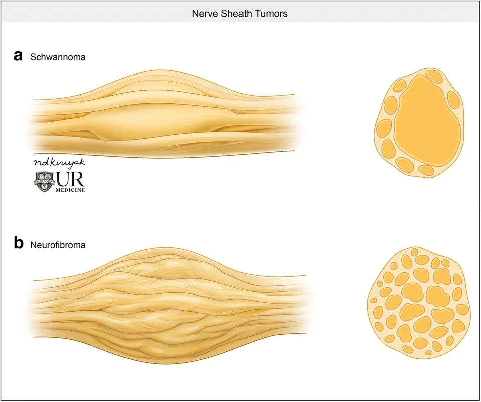
- Schwannoma (also called neurinoma): the most common type. It grows from the cells that insulate the nerve fibres, usually sits on the outside of the nerve and pushes the working fibres aside rather than growing through them. Up to 9 in 10 schwannomas are solitary and occur without a hereditary condition, mainly between the ages of 30 and 60.
- Neurofibroma: grows in between the nerve fibres, so that they run through it. This makes complete removal more difficult. Several neurofibromas, or an extensive "plexiform" neurofibroma, fit with the hereditary condition neurofibromatosis type 1 (NF1).
- Malignant peripheral nerve sheath tumour (MPNST): the malignant form, rare (about 1 to 2 new cases per million people per year). It sometimes develops in an existing plexiform neurofibroma in NF1, sometimes without any prior history.
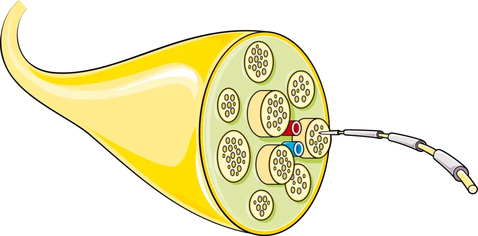
A nerve tumour is therefore something different from an entrapment such as carpal tunnel syndrome, although the symptoms can be similar.
What symptoms does it cause?
- A slowly growing lump, often painless, that can be moved from side to side but not along the length of the nerve
- Tapping on the lump causes tingling or an electric shock in the area of the nerve (Tinel's sign)
- Pain, especially when pressure is put on the lump or at night
- Less often (in fewer than 1 in 5): numbness or loss of strength in the area of the nerve; this points more towards a neurofibroma or a larger tumour
Warning signs: a lump that clearly gets bigger within a few weeks or months, is larger than about 5 cm, causes pain at rest or at night, causes rapidly increasing loss of strength, or develops in someone with neurofibromatosis type 1. These are reasons for prompt further investigation, because a malignant tumour must then be ruled out.
How is the diagnosis made?
Ultrasound is a good first test for a superficial lump: it shows whether the swelling arises from a nerve and can guide a needle sample if needed. MRI is the reference test: it shows the tumour, its relationship to the nerve and the surrounding structures, and helps plan the operation. A CT scan is of little use here.
Important to know: imaging cannot tell a schwannoma from a neurofibroma with certainty, nor always a benign tumour from a malignant one. If malignancy is suspected, especially in NF1, a PET scan is requested: it identifies an MPNST with an accuracy of about 90%. A needle biopsy is avoided when the appearance is typically benign, because it can damage the nerve and adds little; it is considered when a malignant tumour is suspected and the treatment depends on it.
An EMG is rarely needed for the diagnosis, but can be useful to record the function of the nerve before an operation.
Does every nerve tumour need an operation?
No. A small, painless tumour that looks clearly benign and causes no loss of function can safely be monitored with a clinical check-up and an MRI after six to twelve months, and then at longer intervals if the tumour remains stable. Many schwannomas barely grow for years.
An operation is proposed in case of:
- pain or bothersome tingling that limits daily life
- demonstrable growth on successive scans
- numbness or loss of strength that is increasing
- doubt about the nature of the tumour, or warning signs of malignancy
- an explicit wish of the patient after an honest discussion of the pros and cons
For a proven or strongly suspected MPNST, prompt wide removal by a specialised team (sarcoma centre) is needed, often with radiotherapy and sometimes chemotherapy. This care is organised in consultation with oncologists and radiation oncologists and, where appropriate, the patient is referred.
How does the operation go?
The aim is to remove the tumour completely while preserving the working nerve fibres. With a schwannoma this usually works well, because the tumour arises from a single fibre bundle and pushes the other bundles aside.
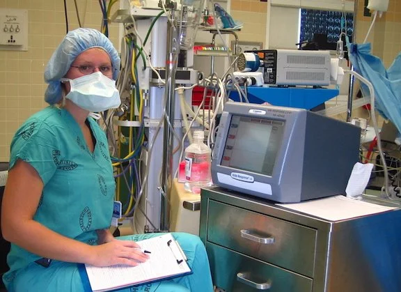
The procedure is done under general anaesthesia and under the operating microscope. The nerve is exposed above and below the tumour. Using a fine electrical stimulator, the nerve fibres on the surface of the tumour are mapped (intraoperative nerve monitoring): this way the covering is opened at the spot where no or as few working fibres as possible run. The tumour is then "shelled out" of its capsule (enucleation) and only the fibre bundle from which it arose is divided, after stimulation has confirmed that it no longer has any function. The surrounding bundles remain intact.
With a neurofibroma, which grows in between the fibres, complete removal without loss of function is not always possible. In that case it is agreed beforehand how much of the tumour will be removed and whether a nerve repair (using a piece of sensory nerve from the lower leg as a bridge) is advisable. This happens rarely, and never unannounced.
Most procedures on the arm or leg are done as day surgery or with one overnight stay. The removed tissue is always examined by the pathologist; that result determines further follow-up.
What are the risks?
Besides the general risks of surgery (bleeding after the operation, infection, a tender scar), the specific risk is new loss of nerve function. In a large series of 182 benign nerve tumours, about 1 in 10 patients had temporary new loss of strength after removal of a schwannoma and just over 1 in 10 had new loss of sensation; in about half of them a residual deficit remained. New nerve pain occurred in fewer than 1 in 20 after a schwannoma and more often after a neurofibroma. The more completely the tumour can be removed without harming the nerve, the better the result; intraoperative monitoring serves exactly that purpose.
A benign tumour that has been completely removed rarely comes back: about 1 in 20 schwannomas and slightly more often with neurofibromas, especially if not everything could be removed or in NF1. Radiotherapy is not needed after complete removal of a benign tumour.
Neurofibromatosis (NF1 and NF2)
In NF1, multiple skin neurofibromas almost always develop, and in about half of patients an extensive plexiform neurofibroma. These are not removed as a matter of routine, only in case of symptoms or growth. The lifetime risk of a plexiform neurofibroma becoming malignant is 8 to 13%; that is why new pain, rapid growth or new loss of function in NF1 is always investigated promptly, with a PET scan if needed. For plexiform neurofibromas that cause symptoms and cannot be operated on safely, there are targeted medicines (MEK inhibitors). These were initially available only for children; since 2025 there has also been a medicine for adults in Europe. They are prescribed in consultation with the (paediatric) oncologist.
In NF2, mainly schwannomas develop, typically of the hearing and balance nerve on both sides, but also on peripheral nerves and in the spinal canal. These patients are followed up by a multidisciplinary team; a peripheral schwannoma is only operated on if it causes symptoms, because there are usually several lesions.
Anyone who has several nerve tumours or who is diagnosed at a young age is referred for genetic counselling.
Frequently asked questions
Is a nerve tumour cancer?
Usually not. The great majority are a benign schwannoma or neurofibroma that grows slowly and does not spread. Malignant nerve sheath tumours are rare and are usually accompanied by warning signs such as rapid growth or pain at rest.
Does a biopsy have to be done first?
Not when the appearance is typically benign: a needle biopsy can damage the nerve and rarely changes the management. If malignancy is suspected, it is considered, usually under imaging guidance and after multidisciplinary discussion.
Is the nerve cut during the operation?
Not with a schwannoma: the tumour is shelled out of the nerve and only the single fibre bundle from which it arose, which no longer has any function, is divided. The nerve keeps working.
What is intraoperative nerve monitoring?
During the operation, the nerve fibres are stimulated with a weak electrical current while the muscles are monitored. This lets us see which fibres are still working and where we can safely open and cut.
Can I just leave the tumour where it is?
Often, yes. A small, painless, stable tumour without loss of function is monitored with an MRI. If it grows, becomes painful or causes loss of function, an operation is discussed again.
How long does recovery take?
The wound heals in two weeks; the scar can remain tender for a few months. Light activities are possible after a few days, heavier loading of the operated limb after four to six weeks, by agreement. Temporary tingling in the area of the nerve is normal and usually decreases over the course of weeks.
Related treatments
All treatments →