
home > treatments > peroneal nerve injury
Peroneal nerve injury (foot drop)
Narrowing of or pressure on the nerve beside the knee, with foot drop
What is a peroneal nerve injury?
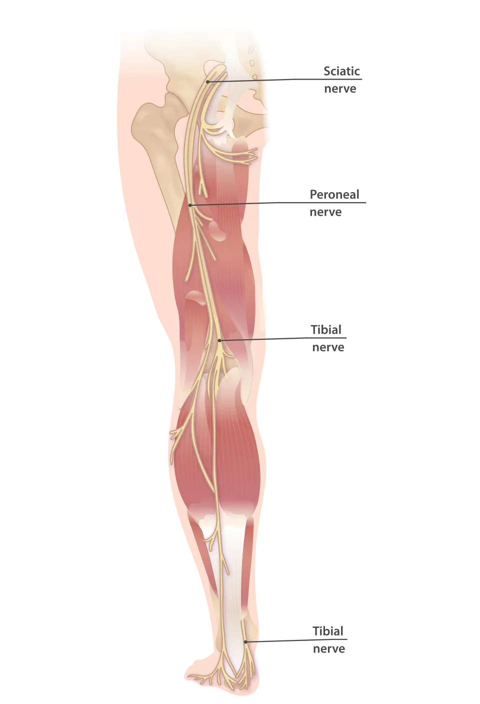
The peroneal nerve (also called the fibular nerve) runs superficially around the head of the fibula on the outer side of the knee, just under the skin. At that spot the nerve is vulnerable to pressure and stretch. It controls the muscles that lift the foot and toes and turn the foot outwards, and it provides sensation to the outer side of the lower leg and the top of the foot.
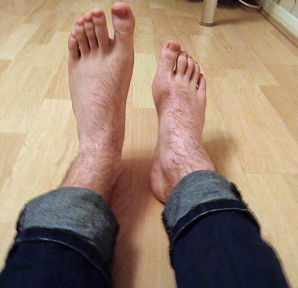
If the nerve becomes trapped or damaged there, foot drop develops: the foot can no longer be lifted and slaps onto the ground when walking. It is the most common nerve entrapment in the leg.
How does it develop?
- External pressure: a habit of crossing the legs, squatting or kneeling for long periods, a plaster cast, a tight knee brace, lying in bed or on an operating table for a long time
- Weight loss: losing a lot of weight quickly (diet, weight-loss surgery, illness, including with weight-loss medication) makes the protective fat pad around the nerve disappear; this is now the most frequently recognised risk factor
- Injury: a fracture or dislocation of the knee, a severe ankle sprain, knee surgery or a knee replacement
- Space-occupying lesions: rarely a cyst from the knee joint pressing on the nerve, or a nerve tumour
- No clear cause: in about half of patients
Diabetes, a generalised nerve disorder (polyneuropathy), excessive alcohol use and smoking make the nerve more vulnerable and recovery slower.
What symptoms does it cause?
- No longer being able to lift the foot or toes; tripping over your own foot, lifting the knee higher when walking (steppage gait)
- Numbness or tingling on the outer side of the lower leg and on the top of the foot, extending to between the big toe and the second toe
- Sometimes pain or an electric feeling on the outer side of the knee when pressure is applied or when the knee is straightened
- Unsteady walking and an increased risk of falling
Foot drop is not always a problem of the nerve at the knee. A disc herniation in the lower back that traps the L5 nerve root can produce exactly the same picture, but then usually with back pain and radiating pain, and with weakness of the hip muscles as well. That distinction determines the treatment and is made at the consultation and with the EMG.
Warning signs: foot drop that comes on suddenly together with severe back pain, weakness in both legs, numbness in the saddle area or problems passing urine or opening your bowels is a reason to see a doctor or go to the emergency department the same day. Foot drop after a knee injury or dislocation also needs to be assessed quickly.
How is the diagnosis made?
The clinical examination shows which muscles are weakened and whether the sensory disturbance matches the area of this nerve. This distinguishes peroneal nerve compression from a problem in the back or a generalised nerve disorder.
The EMG (nerve conduction study) is the most important test. It confirms that the nerve is affected at the level of the head of the fibula and, more importantly, it says something about the prognosis. If the nerve is only "blocked" but the nerve fibres are intact (a so-called conduction block), the chance of recovery is high. If nerve fibres have died off (axonal damage), recovery is slower and sometimes incomplete. Even then, about half of patients still recover well, so a poor EMG does not mean that recovery is ruled out. The EMG is most reliable from three weeks after the start of symptoms.
An ultrasound scan of the nerve is a good, inexpensive first imaging test: it shows a thickened nerve, the exact site of the entrapment and any cyst or tumour. An MRI is requested if there is doubt, if a space-occupying lesion is suspected or to assess the muscles.
Treatment: first without surgery
The first treatment is almost always non-surgical, because a considerable proportion of patients recover spontaneously. In recent Belgian research, about 3 in 10 patients had already recovered within the first three months, before an operation was even considered.
- Taking away the pressure: no longer sitting with crossed legs, no squatting or kneeling, no tight knee brace or compression stocking that presses at the level of the head of the fibula
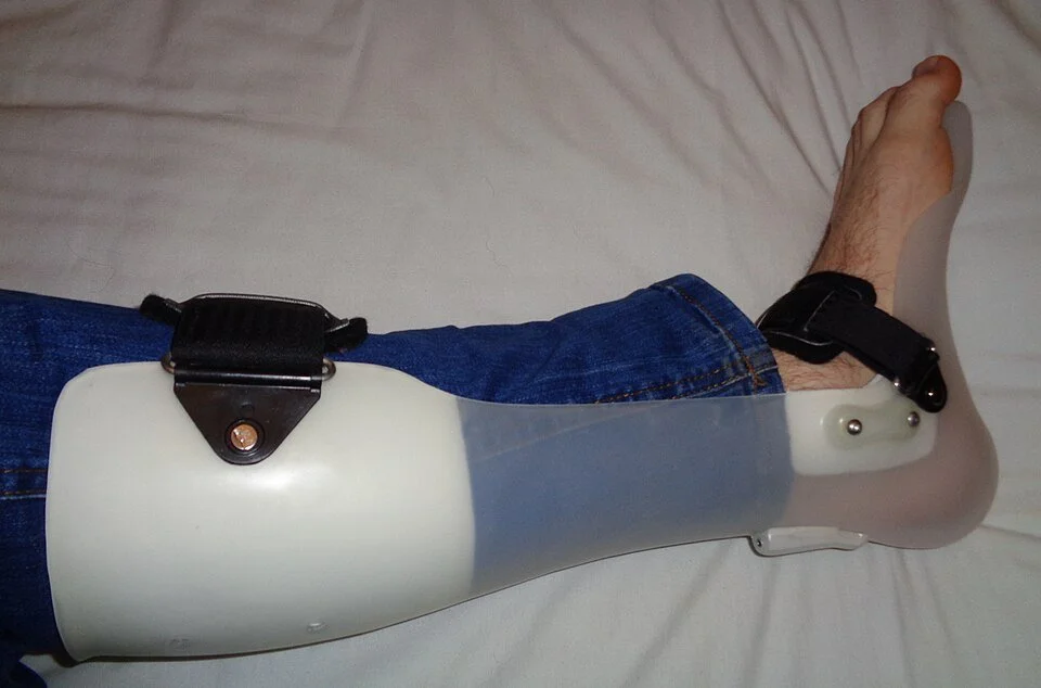
- Ankle-foot orthosis (foot-drop splint): a light splint that lifts the foot when walking. It does not heal the nerve, but it makes safe and easy walking possible and prevents falls and shortening of the calf muscle while the nerve recovers
- Physiotherapy: practising walking and balance, maintaining the strength of the surrounding muscles, stretching the calf muscle so that the ankle stays supple
- Tackling the underlying cause: keeping diabetes well controlled, not letting weight loss happen too quickly, enough protein and vitamins when dieting
When to discuss an operation?
If the foot drop does not recover within six to fourteen weeks, we discuss an operation. To be honest: whether an operation at that point is better than continuing to wait with exercise therapy has not been scientifically proven. A Belgian randomised trial that compared the two (FOOTDROP, 2026) had to stop early because too few patients could take part, partly because of the high rate of spontaneous recovery; among the 26 patients treated, there was no difference in walking distance after nine months, and the patients without surgery recovered faster on average. Large observational series show good results in both groups: about 85 in 100 after an operation and about 80 in 100 without.
That is why the decision depends on your situation: a clear site of entrapment on ultrasound, signs of nerve fibre damage on the EMG without any recovery after three months, a cyst or other lesion pressing on the nerve, or severe pain at the site of the nerve are arguments for surgery. Waiting until after a year is in any case unfavourable: after twelve months of foot drop, the results of an operation are clearly worse.
How does the operation go?
The procedure is called decompression or neurolysis of the peroneal nerve. Through an incision of a few centimetres on the outer side of the knee, just below the head of the fibula, the bands of connective tissue and the muscle edge that are constricting the nerve are divided, so that the nerve lies free along its entire course. Any cyst is removed during the same procedure. The operation usually takes less than an hour, is done under general or regional anaesthesia and usually as day surgery or with one overnight stay. The nerve itself is not cut or moved.
Nerve fibres that were damaged then have to grow back to the muscle, at about one millimetre per day. Recovery of the muscles that lift the foot after an operation can therefore take from a few weeks to many months; in the largest series, the average recovery time was about three months.
What are the risks?
Bleeding after the operation, infection, a tender scar and temporarily reduced feeling around the incision. Rare: injury to the nerve or to a sensory branch, or no recovery despite a successful release, especially when the nerve had been severely damaged for a long time. In the recent Belgian research, 2 in 14 patients who had surgery had a complication, none of them serious. Long-standing loss of sensation often recovers only partially.
How does recovery go?
You may walk from the first day, with the orthosis as long as the foot does not yet lift. The stitches are removed after two weeks; you can drive as soon as you can use your right foot safely, so with right-sided foot drop only once the muscles that lift the foot return. Physiotherapy is continued to train the muscles as soon as they are being activated again. The EMG can be repeated after a few months to document the recovery. If the ability to lift the foot has still not returned after one to two years, there are orthopaedic procedures (tendon transfer) that can improve walking; these are discussed in consultation with the orthopaedic surgeon.
Frequently asked questions
Does foot drop always need an operation?
No. A large proportion recover spontaneously, especially when the cause was external pressure or weight loss. An operation is only discussed if there is no recovery after a few months or if there is a clear cause of entrapment.
Is my foot drop coming from my back or from my knee?
Both are possible and the distinction is important. Back pain with radiating pain, weakness of the hip muscles as well or an abnormal MRI of the back point to a disc herniation; pain and a thickened nerve at the knee and a typical EMG point to the peroneal nerve.
How long does recovery take?
With a pure conduction block, often a few weeks to three months. If nerve fibres are damaged, they have to grow back: about a millimetre per day, so months to more than a year.
I lost a lot of weight quickly and developed foot drop. Is that a coincidence?
No, that is a known cause: the fat pad that protects the nerve at the knee disappears. Recovery in this group is usually good. In the meantime, avoid crossing your legs and squatting.
Can I drive with foot drop?
With right-sided foot drop, driving an ordinary car is not safe, not even with an orthosis. Discuss this with your doctor; sometimes an adapted car or an automatic with left-foot controls is a solution.
What if the nerve does not recover?
Then the orthosis remains a good way of walking safely. At a later stage, an orthopaedic tendon transfer can partly restore the ability to lift the foot.